
Asymptomatic Osteonecrosis of the Trochlea in an Adolescent: A Case Report

Abstract
Introduction
Osteonecrosis, also known as avascular necrosis, aseptic necrosis, or ischemic necrosis, results from a temporary or permanent halt in blood flow to a portion of bone. This lack of blood supply can eventually cause the affected bone to collapse. Osteonecrosis around the elbow is not frequently observed. However, its occurrence in the trochlea known as Hegemann's disease is even rarer. Incidence rates of trochlear osteonecrosis have been reported to vary from 0.27% to less than 0.001% across different studies.
Case presentation
A 14-year-old male presented with severe right shoulder pain and swelling, along with mild right lateral-sided elbow pain due to a fall to the ground. The radiograph of the right shoulder revealed a proximal humeral metaphyseal greenstick fracture. Additionally, the radiograph of the right elbow incidentally revealed osteonecrosis of the distal humeral trochlea. The affected shoulder was immobilized and Conservative management was selected for treating the trochlear osteonecrosis.
Conclusion
Trochlear avascular necrosis is a rare condition that might cause mild discomfort or even be asymptomatic, potentially being diagnosed incidentally through radiographs. Typically, it can be managed with conservative treatment methods.
Introduction
Osteonecrosis, alternatively termed avascular necrosis (AVN), aseptic necrosis, or ischemic necrosis, occurs due to a temporary or permanent interruption of blood flow to a section of bone.
This deprivation of blood supply can lead to the eventual collapse of the affected bone [1]. This condition often manifests as joint pain, bone damage, and reduced function. Commonly affected areas include the ends of long bones like the femur and humerus, as well as regions like the knee's femoral condyles, the tibial plateau, and the small bones in the hands and feet [2,3]. While AVN around the elbow is not frequently observed, its occurrence in the trochlea is even rarer compared to other elbow regions such as the capitellum, radial head, and olecranon [4].
The term osteochondrosis encompasses over 50 various conditions that affect the developing skeleton. In 1951, Dr. Gerd Hegemann documented the radiographic alterations observed in the humeral trochlea of young adults. Hence, osteochondrosis specifically affecting the humeral trochlea is referred to as Hegemann's disease [5].
Hegemann's disease can arise from either traumatic or non-traumatic causes. Instances involving trauma often involve elbow contusions or fractures as contributing factors [6]. However, Osteonecrosis commonly develops in individuals who have certain risk factors, including high-dose corticosteroid therapy, excessive alcohol consumption, injury, malignancy, systemic lupus erythematosus, and hematologic disorders like sickle cell disease along with certain Infectious causes [1,7].
Osteonecrosis of the trochlea is an extremely rare condition affecting the lower end of the humerus. Incidence rates have been reported to vary from 0.27% to less than 0.001% across different studies [6,8].
This report presents a rare case of trochlear osteonecrosis in an adolescent. All of the references cited in this report were evaluated for eligibility [9].
Case Presentation
Patient information
A 14-year-old male was brought to the emergency department of our hospital with severe right shoulder pain and swelling, along with mild right lateral-sided elbow pain. These symptoms had started approximately two hours after he fell to the ground. Before the fall, the patient did not complain of any pain or limitation of range of motion in either joint. The patient's parents reported two previous traumas. The first, at the age of eleven, involved a fall on an outstretched hand, resulting in mild elbow pain for approximately three days, which resolved without medical intervention. The second incident occurred one year prior in a road traffic accident, resulting in a right distal tibial greenstick fracture. However, there were no concurrent upper limb complaints during this episode, and the fracture was managed conservatively with long leg casting.
Clinical findings
The patient had severe tenderness over the proximal humerus with limitation of shoulder range of motion due to pain and mild swelling. He had also complained about mild right lateral-sided elbow tenderness with a normal elbow range of motion and no elbow deformity was noted with the normal neurovascular examination of that limb.
Diagnostic assessment
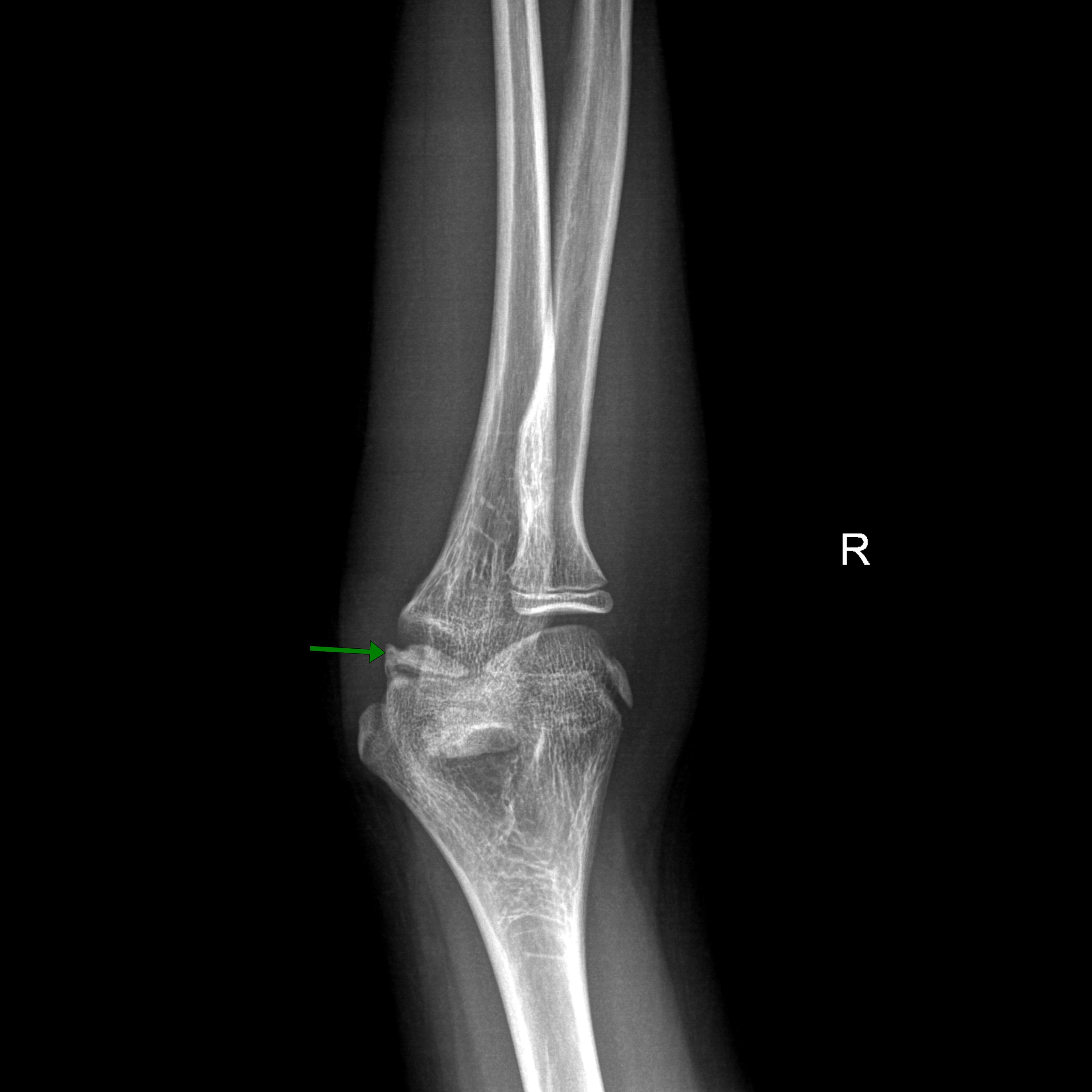
The radiograph of the right shoulder revealed a proximal humeral metaphyseal greenstick fracture, (figure. 1). Additionally, the radiograph of the right elbow incidentally revealed osteonecrosis of the distal humeral trochlea, with no other superimposed findings noted. Notably, the carrying angle was measured at 12 degrees in valgus.
Therapeutic intervention
A sling and swathe were applied to immobilize the affected shoulder, and the patient was provided analgesics. Conservative management was selected for treating trochlear osteonecrosis, which involved incorporating a range of motion exercises after the proximal humerus fracture had fully healed. Close follow-up was arranged to monitor his progress. Subsequently, he was discharged from the hospital.
Follow-up and Outcome
During the follow-up, the patient had no complaints regarding his elbow.
Discussion
The exact causes of Hegemann's disease remain unidentified. Nevertheless, various traumatic and non-traumatic factors have been conclusively associated with trochlear osteonecrosis. These include acute or past trauma such as fractures, persistent repetitive microtrauma, and contusions. Additionally, in some cases, the condition may arise without an identifiable cause, being classified as idiopathic [8,10,11]. However, certain risk factors have been associated with osteonecrosis such as corticosteroid therapy, alcohol consumption, bone injuries, systemic conditions such as malignancy, lupus erythematosus, sickle cell disease, Gaucher's disease, Caissons disease, gout, vasculitis, osteoarthritis, osteoporosis, radiation therapy, chemotherapy, and organ transplantation, particularly renal transplants [7]. Rarely, infections such as HIV and meningococcemia leading to disseminated intravascular coagulation have been associated [12,13]. Nonetheless, a notable proportion of cases remain idiopathic [7]. In this study, the patient had a history of two previous traumas, followed by a recent fall to the ground.
The ossification center of the trochlear epiphysis typically becomes visible after the age of five, progressing in development between 8 and 13 years in boys. Fusion with the humeral metaphysis occurs between the ages of 13 and 16. [8]. Two vessels enter the posterior aspect of the lateral humeral condyle and traverse an extended path through the lateral condylar ossification center, ultimately reaching the lateral section of the trochlea. The trochlea itself is nourished by these lateral vessels, along with a distinct vessel that permeates the medial, nonarticular portion of the trochlea [6]. The presence of these two blood supplies gives rise to a watershed area within the trochlear groove. Disruption of this distinctive blood supply can occur during the injury, as well as during closed or open reduction maneuvers, or internal fixation procedures [6,14].
Trochlear AVN can manifest either partially or entirely. In Type A cases, where there is partial involvement, the apex or lateral segment of the trochlear medial crista is typically affected. Patients in this category typically show no symptoms and do not exhibit angular deformities. Radiologically, they display a central deficiency in the distal humeral epiphysis. Conversely, in Type B cases, where there is complete involvement, the entire trochlear metaphysis is affected. These patients often experience a gradual onset of elbow varus deformity and a notable reduction in range of motion [8].
According to Schumacher et al., Hegemann's disease progresses through five distinct stages as observed on radiographs [15]. In Stage 1, there is an initial decrease in density followed by plaque-like sclerosis in the center of epiphyseal ossification. Stage 2 is characterized by a decrease in size and increased condensation of the ossification center. In Stage 3, loosening occurs along with the emergence of new ossification. Stage 4 is marked by regeneration and enlargement of the ossification center. Finally, Stage 5 represents the ultimate stage, which may involve either complete or partial recovery [11,16].
Uhrmacher et al. were the pioneers in identifying Hegemann's disease in two children aged 7 and 9 years. The primary symptoms observed were swelling and limited range of motion in the elbow [17]. In the current case, the patient presented with significant discomfort characterized by severe right shoulder pain and swelling attributed to a proximal humeral metaphyseal greenstick fracture incurred from a fall, accompanied by mild discomfort localized to the right lateral aspect of the elbow. The elbow exhibited a normal range of motion, with no observable deformity noted. Notably, preceding the recent accident, the patient had been asymptomatic for trochlear AVN.
Hegemann's disease is frequently identified through radiographic examination months or even years following trauma, leading to potential confusion with a condition known as fishtail deformity. This deformity, uncommonly encountered, typically arises as a complication after a distal humeral fracture during childhood [5]. Hegemann’s disease was initially identified before the availability of computed tomography (CT) scans or magnetic resonance imaging (MRI) techniques. Consequently, the fishtail deformity might have been considered a subsequent stage of Hegemann’s disease, which is typically benign following a mild vascular disorder. However, complete AVN could potentially develop following traumatic incidents. Another perspective suggests that Hegemann’s disease could represent a benign, self-limiting phase of the fishtail deformity after unrecognized injury or repetitive micro-trauma. Characterized by irregularity of the trochlea and sclerosis, Hegemann’s disease presents distinct clinical features [5]. However, Beyer et al. showed that trochlear aseptic necrosis exhibits a low-intensity signal on T1-weighted MRI images. They also emphasized MRI's utility in diagnosing Hegemann's disease and confirming recovery [11]. In the current report, the radiograph of the right elbow incidentally revealed osteonecrosis of the distal humeral trochlea, with no other superimposed findings noted.
The objective of treating AVN is to enhance the functionality of the affected joint, prevent further deterioration of the bone, and secure the survival of both bone and joint structures. Identifying and addressing the underlying cause of AVN is imperative whenever feasible [7]. A review conducted by Claessen et al. observed that all eight documented cases of Hegemann disease underwent conservative treatment, involving rest and modifications in activity. Among the five patients with recorded clinical progress, four experienced complete alleviation of pain following conservative management, while the fifth patient continued to experience intermittent pain [5]. However, surgical treatment options such as arthroscopic debridement, core decompression, vascularized bone grafting, and bone reconstruction are recommended when symptoms persist and signs of collapse become apparent [1]. In the present case, the affected shoulder was immobilized using a sling and swathe, and the patient received pain relief medication. Conservative treatment was chosen for trochlear osteonecrosis, including the range of motion exercises once the proximal humerus fracture had healed.
Conclusion
Trochlear AVN is a rare condition that might cause mild discomfort or even be asymptomatic, potentially being diagnosed incidentally through radiographs. Typically, it can be managed with conservative treatment methods.
Declarations
Conflicts of interest: The author(s) have no conflicts of interest to disclose.
Ethical approval: Not applicable.
Patient consent (participation and publication): Written informed consent was obtained from the parent of the patient for publication.
Funding: The present study received no financial support.
Acknowledgements: None to be declared.
Authors' contributions: AKG was a significant contributor to the conception of the study and the literature search for related studies. SSR, RJR, DQH, BJR and PHR were involved in the literature review, the study's design, and the critical revision of the manuscript, and they participated in data collection. HAN and KKM were involved in the literature review, study design, and manuscript writing. SHT was the radiologists who performed the assessment of the case. HAN and AKG confirm the authenticity of all the raw data. All authors approved the final version of the manuscript.
Use of AI: AI was not used in the drafting of the manuscript, the production of graphical elements, or the collection and analysis of data.
Data availability statement: Not applicable.
References
- Agarwal R, Gupta R, Singh S, Gupta K, Kudesia M. Avascular necrosis of humeral head in an elderly patient with tuberculosis: a case report. Journal of Medical Case Reports. 2008; 2:1-3. doi:10.1186/1752-1947-2-361
- Franceschi F, Franceschetti E, Paciotti M, Torre G, Samuelsson K, Papalia R. Surgical management of osteonecrosis of the humeral head: a systematic review. Knee Surgery, Sports Traumatology, Arthroscopy. 2017;25(10):3270-8. doi:10.1007/s00167-016-4169-z
- Mankin HJ. Nontraumatic necrosis of bone (osteonecrosis). New England Journal of Medicine. 1992;326(22):1473-9. doi:10.1056/NEJM199205283262206
- Bakarman K, Alghnimei N, Borai S, Alfadhil R. Bilateral trochlear avascular necrosis: A case report and brief review of the literature. International Journal of Surgery Case Reports. 2021;86:106303.doi:10.1016/j.ijscr.2021.106303
- Claessen FM, Louwerens JK, Doornberg JN, van Dijk CN, van den Bekerom MP, Eygendaal D. Hegemann's disease and fishtail deformity: aetiopathogenesis, radiographic appearance and clinical outcome. Journal of Children's Orthopaedics. 2015;9(1):1-8. doi:10.1007/s11832-014-0630-z
- Hamilton DA, Kalra K. Trochlear osteonecrosis after a nonoperative lateral humeral condyle fracture in a child. JAAOS Global Research & Reviews. 2020 May 1;4(5):e19. doi:10.5435/JAAOSGlobal-D-19-00101
- Assouline-Dayan Y, Chang C, Greenspan A, Shoenfeld Y, Gershwin ME. Pathogenesis and natural history of osteonecrosis. InSeminars in arthritis and rheumatism (Vol. 32, No. 2, pp. 94-124). WB Saunders.
- Bakarman K, Alghnimei N, Borai S, Alfadhil R. Bilateral trochlear avascular necrosis: A case report and brief review of the literature. International Journal of Surgery Case Reports. 2021;86:106303. doi:10.1016/j.ijscr.2021.106303
- Kakamad FH, Abdalla BA, Abdullah HO, Omar SS, Mohammed SH, Ahmed SM, et al. Lists of predatory journals and publishers: a review for future refinement. European Science Editing. 2024;50: e118119. doi:10.3897/ese.2024.e118119
- Assad SK, Sabah M, Kakamad FH, Salih AM, Salih RQ, Mohammed SH, et al. Avascular necrosis of femoral head following COVID-19 infection. Annals of Medicine and Surgery. 2023;85(9):4206-10. doi:10.1097/MS9.0000000000001098
- Beyer WF, Heppt P, Glückert K, Willauschus W. Aseptic osteonecrosis of the humeral trochlea (Hegemann's disease). Archives of orthopaedic and trauma surgery. 1990;110:45-8. doi:10.1055/s-0034-1371835
- Qaqish RB, Sims KA. Bone disorders associated with the human immunodeficiency virus: pathogenesis and management. Pharmacotherapy: The Journal of Human Pharmacology and Drug Therapy. 2004;24(10):1331-46. doi:10.1592/phco.24.14.1331.43150
- Campbell WN, Joshi M, Sileo D. Osteonecrosis following meningococcemia and disseminated intravascular coagulation in an adult: case report and review. Clinical infectious diseases. 1997;24(3):452-5. doi:10.1093/clinids/24.3.452
- Kimball JP, Glowczewskie F, Wright TW. Intraosseous blood supply to the distal humerus. The Journal of hand surgery. 2007;32(5):642-6. doi:10.1016/j.jhsa.2007.02.019
- Schumacher R, Müller U, Schuster W. Rare localisation of osteochondrosis juvenilis (author's transl). Der Radiologe. 1981;21(4):165-74.
- Beaty JH, Kasser JR. Rockwood and wilkin's fractures in children. InRockwood and Wilkin's fractures in children 2010 (pp. 1076-1076).
- Uhrmacher F. Uber Osteochondritis deformans juvenilis des Ellenbogengelenkes. Z Orthop Chir. 1933;59:398-411.

This work is licensed under a Creative Commons Attribution 4.0 International License.