Vol 3, No 2 (2025): Current Issue (Volume 3, Issue 2), 2025
Editorial
Public-Private Partnerships: A Pathway to Health Equity in Developing Countries
Dahat A. Hussein
In the face of mounting healthcare challenges, developing countries grapple with resource constraints, underfunded health systems, and a growing burden of infectious and non-communicable diseases. The question is no longer whether healthcare systems can sustain themselves under these pressures—the evidence says they cannot—but how they can adapt and evolve to meet these needs. One promising approach lies in establishing and strengthening public-private partnerships (PPPs) [1].
PPPs are collaborations in which governments and private entities collaborate to achieve common health goals, leveraging the strengths of both sectors. The public sector offers reach, regulatory frameworks, and an understanding of population health needs, while the private sector contributes innovation, efficiency, and financial resources. Together, they can address healthcare access, quality, and affordability gaps [1].
In many developing countries, healthcare access remains unequal. Rural and underserved populations often lack essential services, while urban areas face overcrowded and overburdened public hospitals. By pooling resources and expertise, PPPs can deliver sustainable solutions, such as building health facilities in remote areas, funding community health programs, or providing cutting-edge technology and training to public hospitals. Initiatives like the African Access Initiative have brought affordable medicines and diagnostic tools to low-income countries by fostering collaborations between governments and pharmaceutical companies. These partnerships ensure the availability of life-saving medications for conditions like cancer and diabetes [2]. In countries like India, PPPs have been instrumental in building state-of-the-art hospitals in rural areas, combining public funding with private expertise. For example, the Karnataka state government’s partnership with private hospitals expanded tertiary healthcare access to previously underserved populations [3]. Partnerships with multinational corporations and NGOs have facilitated training for healthcare professionals, enabling them to provide higher-quality care. In Kenya, PPPs have focused on improving maternal and child health outcomes through community-based education programs [4], in 2010, the Kurdistan Regional Government (KRI) asked for assistance from the RAND Corporation, a U.S.-based nonprofit policy think tank specializing in research and development across various fields, to support the reform of the healthcare system in the Kurdistan Region of Iraq. The primary goal of the reform was to establish a health system that could efficiently provide high-quality services to all individuals, with the support of Public-Private Partnerships (PPPs) [5].
Despite their promise, PPPs are not without challenges. Critics argue that these arrangements can lead to the privatization of public health assets or create inequities in service delivery. For instance, poorly regulated PPPs may prioritize profitability over the needs of vulnerable populations, leaving rural or low-income communities underserved. Transparency, accountability, and robust legal frameworks are essential to mitigate these risks. Governments must ensure that PPP agreements prioritize public health outcomes with clear success metrics and oversight mechanisms.
As the global health landscape evolves, developing countries have an opportunity to harness PPPs as a tool for resilience and equity. By fostering inclusive partnerships that align public and private incentives, countries can mobilize resources, improve service delivery, and ensure sustainable healthcare financing.
The need for innovation and collaboration in healthcare has never been more urgent. Governments, private entities, and civil society must work together to create partnerships that prioritize the well-being of all citizens, particularly the most vulnerable. With the right frameworks, public-private partnerships can move beyond being a stopgap solution and become a cornerstone of sustainable health systems.
Let us embrace the potential of PPPs and work toward a future where healthcare is a right, not a privilege, for everyone, everywhere.
Original Articles

Desmoid-Type Fibromatosis of The Breast: A Case Series
Ari M. Abdullah, Zuhair D. Hammood, Lana R.A. Pshtiwan, Rawa M. Ali, Abdulwahid M. Salih, Hadeel...
Abstract
Introduction
Desmoid-type fibromatosis (DTF), also called aggressive fibromatosis, is a rare, benign, locally aggressive condition. Mammary DTF originates from fibroblasts and myofibroblasts within the breast tissue, representing 0.2% of all breast tumors. This study aims to present and discuss the clinical presentation and management of seven cases of breast DTF.
Methods
This single-center case series was conducted at the breast clinic of Smart Health Tower in Sulaymaniyah, Iraq. It included all patients diagnosed as breast DTF by histopathological examination, with those lacking complete data excluded. The patients were treated and managed between January 2021 and August 2024.
Results
This study involved seven female patients with a mean age of 35.29 ± 14.29 years. Clinically, six of them (85.71%) presented with a non-tender palpable breast mass, while one patient (14.28%) reported breast pain. Ultrasound revealed hypoechoic lesions in all cases. The average size of the masses was 29.43 ± 17.26 mm. All patients underwent wide local excision of the breast mass. Histopathological examination confirmed the diagnosis of DTF in all cases.
Conclusion
Diagnosing a desmoid tumor of the breast can be difficult, as it can mimic breast carcinoma. The Wide local excision is often the preferred treatment to prevent future recurrences.
Introduction
Desmoid-type fibromatosis (DTF), also known as aggressive fibromatosis, is a rare benign condition that can develop in various body parts, the most common being the extremities, abdominal wall, and intra-abdominal cavity [1]. Mammary DTF arises from fibroblasts and myofibroblasts within the breast tissue. It accounts for 0.2% of all breast tumors [1,2]. Although it does not metastasize, it is known for its local aggressiveness and high recurrence rate [2].
The term "desmoid" was introduced by Mueller in 1838, deriving from the Greek word "desmos," which refers to its tendon-like consistency. However, MacFarlane first described the disease in 1832 [3].
The DTF may occur sporadically or develop following surgical trauma, the implantation of silicone breast implants, or in association with Gardner's syndrome. While most cases of breast fibromatosis are reported in females, it can also occur in males [1]. It commonly affects individuals between 15 and 60 years of age, with the highest incidence occurring in the third and fourth decades of life [4].
Unpredictable and invasive growth patterns characterize the DTF. The tumor often proliferates during the early stages and may also accelerate due to pregnancy or hormonal changes. After this initial growth phase, many patients experience an extended period where the tumor remains stable [5].
Due to their rarity, desmoid tumors present a diagnostic and therapeutic challenge, as they often initially resemble breast carcinoma and have a high tendency for recurrence [6]. This study aims to describe and discuss the presentation and management of seven cases of breast DTF. All the references cited in this study were evaluated for eligibility [7].
Methods
Study design and setting
This single-center case series was conducted at the breast clinic of Smart Health Tower (Sulaymaniyah, Iraq). The patients were treated and managed between January 2021 and August 2024.
Participants
The study included all patients with confirmed breast DTF based on histopathological examination, while those with incomplete data were excluded.
Data collection
Data were collected from the hospital’s registry, including demographic details, clinical presentation, physical examination, medical history, breast ultrasound and mammography findings, core needle biopsy (CNB) and fine needle aspiration cytology (FNAC) results, histopathological examination (HPE), tumor size, magnetic resonance imaging (MRI) and computed tomography (CT) scan findings, types of surgeries performed, postoperative complications, follow-up duration, and recurrence.
Intervention
All patients underwent surgery under general anesthesia with the supine position following skin preparation and disinfection. The procedures involved making elliptical, radial, and semicircular incisions over the breast where the mass was suspected. After the skin was incised, a wide local excision (WLE) of the mass was performed. In one case, the mass extended into the chest wall muscles and involved two ribs, which were also excised. The long thoracic and thoracodorsal nerves were preserved in all cases. Lymph nodes from levels I, II, and III were removed. Hemostasis was achieved, a Redivac drain was inserted for each patient, and a chest tube drain was placed for one patient. The surgical site was then closed in layers.
Histopathological examination and preparation
Regarding pathologic examination, the specimens were fixed in 10% neutral buffered formalin for 24 hours prior to grossing. After that, the specimens were examined systemically, with appropriate sections taken from the tumors and the margins. The resultant blocks were then processed with the Sakura Histo-Tek VP1 automated processor using a standard 11-hour processing protocol through alcohol, xylene, and paraffin. Following embedding in paraffin and trimming, the blocks were sectioned onto regular glass slides, kept in an oven overnight, and then stained manually for hematoxylin and eosin (H&E) using Gill II hematoxylin. The slides were then dried, and coverslips were applied.
For immunohistochemistry, the paraffin blocks were sectioned onto charged glass slides and kept in an oven overnight. Antigen retrieval was achieved through boiling using the Dako PT Link with a solution of pH 6 or 9, depending on the target antibody. The slides were then washed with buffer solution and welled using the Dako Pen, followed by blocking endogenous peroxidase using hydrogen peroxide. The primary antibodies were then applied, followed by the secondary antibody (horseradish peroxidase) and the chromogen (diaminobenzidine). Counterstaining was achieved using hematoxylin Gill II, followed by drying and applying coverslips.
Data analysis
The data were collected using an Excel spreadsheet (Microsoft Excel 2021). They were analyzed qualitatively with Statistical Package for the Social Sciences (SPSS) software (version 27.0) and presented as mean, range, frequencies, and percentages.
Results
This study included seven female patients with an average age of 35.29 ± 14.29 years, ranging from 18 to 61 years. All of the patients were unemployed, and five of them were married (71.43%). Five patients' medical histories were unremarkable (71.43%), while two patients (28.57%) had hypothyroidism. Three patients (42.86%) had previously undergone different types of breast surgeries. None of the patients had a family history of breast cancer.
Among the clinical presentations, six patients (85.71%) had a non-tender palpable breast mass, while one patient (14.28%) experienced breast pain. Ultrasound showed hypoechoic lesions in all patients with BI-RADS 4 or 5 in four patients (57.14%). The average mass size was 29.43 ± 17.26 mm, ranging from 15 mm to 70 mm. In three cases (28.57%), MRI was used, revealing either heterogeneous or homogeneous masses. In one case (14.28%), a CT scan detected a mass in the chest wall with muscle invasion. A CNB was performed in 5 patients (71.43%), and FNAC of the axillary lymph node, which indicated benign lymphoid tissue, was performed in one patient (14.28%).
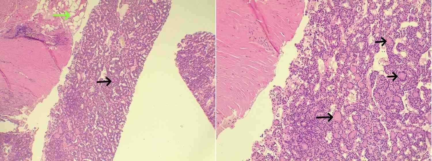
Mammography was performed in only one case (14.28%), showing scattered fibro-glandular density and benign calcification. All patients underwent WLE of the breast mass. In one case (14.28%), surgery required rib excision due to chest wall invasion in a recurrent desmoid tumor. The HPE in all cases, along with immunohistochemistry in 5 cases, confirmed the diagnosis of DTF, with no evidence of lymph node metastasis (Fig. 1). The mean follow-up period was 1.51 ± 0.98 years, ranging from 0.7 to 3 years, with no reported recurrences (Table 1).
Case # |
Age (year)/Gender |
Occupation | Marital status | Medical history | Surgical history | Presentation | Mass size, mm (US), BI-RADS | MMG, BI-RADS | MRI | CT scan | HPE of CNB | FNAC of axillary LN | Operation | Post op. comp | Follow-up | Rec. |
1st |
45/F |
Unemployed |
Married |
Hypothyroidism |
WLE of breast mass |
Rt breast mass |
16 x 26 mm, U2 |
N/A |
N/A |
A 47 x 30 mm irregular mass on the right chest wall invades the pectoralis and contacts the intercostal muscle. |
Fibromatosis |
N/A |
Rt breast mass WLE with the removal of two ribs, achieving clear margins due to a recurrence of fibromatosis after two years. |
No |
2.9 yr |
No |
2nd |
61/F |
Unemployed |
Married |
DM, HTN, IHD |
spine surgery, Interventional endoscopy, PCI |
Rt breast mass |
15 x 9 mm, U5 A3 |
fibro glandular density, M2 |
N/A |
N/A |
Fibromatosis |
Benign |
Rt breast mass WLE |
No |
3 yr |
No |
3rd |
37/F |
Unemployed |
Married |
Negative |
Rt breast surgery |
Rt breast pain |
42 x 12 mm, U3 |
N/A |
Circumscribed oval mass at 2 o’clock, 35 x 22 mm, with uniform enhancement and heterogeneous fibro glandular tissue. MR-3. |
N/A |
Spindle cell neoplasm |
N/A |
Rt breast mass WLE |
No |
1 yr |
No |
4th |
34/F |
Unemployed |
Married |
Negative |
C/S, left breast operation |
Lt breast mass |
70 x 40 mm, U4 |
N/A |
N/A |
Lt breast mass, 75 x 45 mm, invading the 4th to 6th intercostal cartilages and pectoralis major, suggestive of recurrent desmoid tumor. |
Low-grade spindle cell neoplasm, recurrent desmoid type fibromatosis. |
N/A |
Lt breast mass WLE due to fibromatosis recurrence after two years. |
No |
1 yr |
No |
5th |
29/F |
Unemployed |
Married |
Negative |
Two C/S |
Lt breast mass |
20 x 10 mm, U3 |
N/A |
Heterogeneous, spiculated enhancement, 45 x 35 mm, with fibro glandular tissue and moderate background enhancement. MR-4. |
N/A |
N/A |
N/A |
Lt breast mass WLE |
No |
1 yr |
No |
6th |
24/F |
Unemployed |
Single |
Negative |
Negative |
Rt breast mass |
50 x 20 mm, U4a |
N/A |
N/A |
N/A |
N/A |
N/A |
Rt breast mass WLE |
No |
1 yr |
No |
7th |
18/F |
Unemployed |
Single |
Hypothyroidism |
Appendectomy |
Rt breast mass |
70 x 40 mm, U4a |
N/A |
Large circumscribed oval mass, 122 x 90 mm, with heterogeneous enhancement, showing fibro glandular tissue and mild background enhancement. MR-3. |
N/A |
Low-grade spindle cell neoplasm |
N/A |
Rt breast mass WLE |
No |
7 mo |
No |
Discussion
DTF is an intermediate soft tissue tumor marked by clonal fibroblastic proliferation originating in the deep soft tissues. This tumor tends to infiltrate surrounding tissues and has a high local recurrence rate but cannot metastasize [8,9]. They can originate primarily from breast tissue or secondarily from the pectoralis major muscle [1]. This condition typically impacts individuals aged 15 to 60 years, with the highest prevalence observed in the third and fourth decades of life [4]. It is approximately twice as common in women as in men [10]. In this study, the average age of the patients was 35.29 ± 14.29 years, ranging from 18 to 61 years, and all were female.
The exact cause of DTF remains unclear. However, associations with Gardner syndrome, prior trauma, and surgery have been documented [6]. Additionally, while silicone implants are mentioned in the literature as a potential cause, intraoperative trauma is considered more likely to be the primary factor [6].
Clinically, DTF of the breast presents with a wide range of manifestations. It is commonly described as a suspicious, mobile, firm, and painless nodule. However, cases of skin retraction and nipple retraction have also been reported in the literature [6]. Lorenzen et al. described 14 cases of breast DTF in their case series, with all patients presenting with a palpable breast mass [6]. Similarly, in the current study, 85.71% of the patients exhibited a painless breast mass, while only one patient reported breast pain instead of a breast mass.
Relying solely on imaging for diagnosing breast DTF is often inadequate due to its invasive characteristics, which can frequently lead to misdiagnosis as BI-RADS 4 or 5 [10]. On ultrasound, desmoid tumors typically appear as hypoechogenic, ill-defined masses [1]. Mammographically, they present as spiculated masses and, in rare cases, may show calcified deposits [1]. MRI is the most effective imaging technique for assessing the extent of the tumor, particularly when the chest wall is involved [11]. On MRI, desmoid tumors may appear as ill-defined hypointense or isointense masses on T1-weighted images and hyperintense masses on T2-weighted images [12]. In the present study, ultrasound showed hypoechoic lesions in each patient, and ultrasound showed BI-RADS 4 or 5 in 57.14%. Mammography was performed for one patient, showing scattered fibro-glandular density with benign calcifications. MRI was done for three patients, revealing either homogeneous or heterogeneous enhancing masses and varying sizes of fibro-glandular tissue. Additionally, CT scans of one case showed a chest wall mass with invasion of the pectoralis muscle.
Diagnosing a desmoid tumor of the breast can be challenging both clinically and radiologically, as it may resemble breast carcinoma [1]. A definitive diagnosis is only possible through HPE of the lesion. The tumor appears macroscopically as a grayish-white, firm, irregular nodular mass [1]. Microscopically, it is characterized by spindle cells with varying amounts of collagen fiber deposition [12]. Histopathologically, desmoid tumors must be distinguished from scar tissue, fibrosarcoma, or fibromatosis-like metaplastic spindle cell tumors [12].
Immunohistochemical analysis, particularly of preoperative biopsies, can aid in diagnosing desmoid fibromatosis. β-Catenin is a key immunohistochemical marker, showing nuclear expression in sporadic and familial cases in up to 80% of patients [6]. Mutations in the β-catenin gene (CTNNB1) are found in most cases of sporadic DTF, leading to the accumulation of this oncoprotein in the nucleus [6]. Elevated β-catenin levels activate the WnT signaling pathway, contributing to tumor development [6]. However, nuclear β-catenin staining is not specific to breast desmoid fibromatosis; it is also occasionally observed in spindle cell carcinomas, 23% of metaplastic breast carcinomas, and up to 93% of benign phyllodes tumors [13].
The treatment of breast desmoid tumors remains controversial due to the limited data available, given the low incidence of the disease [14]. Various approaches may be employed, including surgery, radiotherapy, chemotherapy, and hormonal therapy. Radical surgical excision is generally considered the treatment of choice [1]. However, complete excision may be avoided in carefully selected patients, especially when the surgery could result in poor functional or cosmetic outcomes, such as in cases where the lesion is superficial or subareolar and may require the removal of the nipple-areolar complex [14]. For some women, mastectomy may be recommended if they experience multiple recurrences, have a large tumor, or face difficulties in obtaining a histological diagnosis [15]. This study chose a comprehensive WLE as a preventive measure to avoid any potential future recurrences.
Breast DTF recurrence is common, with a prevalence of 18-29% over 3-6 years. There is also a risk of involvement with thoracic muscles and ribs [15]. WLE can help prevent or reduce the risk of recurrence [1]. For tumors that are unresectable or require extensive surgical procedures, such as major chest wall resection, radiotherapy may be considered as an alternative treatment option [16]. In this study, no recurrences were observed during the follow-up period. The mean follow-up duration was 1.51 ± 0.98 years, ranging from 0.7 to 3 years.
Systemic therapy can be employed for unresectable or recurrent desmoid tumors, as well as for patients who cannot tolerate surgical treatment or when the tumor involves the chest wall [1]. This therapy includes non-cytotoxic approaches, such as hormonal therapy with Tamoxifen alone or in combination with nonsteroidal anti-inflammatory drugs, as well as cytotoxic approaches like chemotherapy [1]. In the current study, one patient underwent a WLE and the removal of two ribs due to an irregular mass on the right chest wall that invaded the pectoralis muscle and contacted the intercostal muscle. A limitation of this study is the short follow-up duration, which may not be long enough to identify potential recurrences.
Conclusion
Diagnosing a desmoid tumor of the breast can be difficult, as it can mimic breast carcinoma. The WLE is often the preferred treatment to prevent future recurrences.
Declarations
Conflicts of interest: The author(s) have no conflicts of interest to disclose.
Ethical approval: The study's ethical approval was obtained from the scientific committee of the Kscien Organization for Scientific Research.
Patient consent (participation and publication): Verbal informed consent was obtained from patients for publication.
Source of Funding: Smart Health Tower
Role of Funder: The funder remained independent, refraining from involvement in data collection, analysis, or result formulation, ensuring unbiased research free from external influence.
Acknowledgements: None to be declared.
Authors' contributions: AMS and AMA were major contributors to the conception of the study, as well as to the literature search for related studies. HAN, MKA and AAQ were involved in the literature review, study design, and writing the manuscript. ZDH, HAY, SHH, SOK, FHK and HAS were involved in the literature review, the design of the study, the critical revision of the manuscript and the processing of the figures. FHK and AMS confirm the authenticity of all the raw data. RMA was the pathologist who performed the histopathological diagnosis. LRAP was the radiologist who performed the assessment of the case. All authors have read and approved the final manuscript.
Use of AI: AI was not used in the drafting of the manuscript, the production of graphical elements, or the collection and analysis of data.
Data availability statement: Note applicable.

Arteriovenous Fistula Creation for Hemodialysis in Patients with End-Stage Renal Disease with and Without Surgical Drain: A Randomized Control Trial
Lokish S. Jaswel, Narayan Oste, Satish Vaidy
Abstract
Introduction
Failure of an arteriovenous fistula (AVF) disrupts hemodialysis access and reduces the available area for future access. Preventive interventions are necessary to avoid AVF failure. This study evaluates the impact of surgical drainage during AVF creation for hemodialysis in patients with end-stage renal disease (ESRD).
Methods
This single-center, phase II, randomized controlled trial was conducted from June 2020 to June 2023. Ninety-four patients were randomly assigned into two groups: Group A (with a surgical drain) and Group B (without a drain). Patients were followed for six months post-surgery. The primary outcome was AVF primary patency, and secondary outcomes included postoperative complications.
Results
The average age of participants was 63.7 years, with 50 male patients. The most common cause of renal failure was glomerular disease (29.8%), and most AVFs were located on the left side (57.4%). Brachiocephalic AVFs were the most frequent type (70.2%). Postoperative hematoma was more common in Group B (42.6%) than in Group A (17%) (P = 0.007). The primary patency rate at six months was higher in Group A (87.2%) compared to Group B (76.6%), though the difference was not statistically significant (P = 0.180).
Conclusion
The use of surgical drainage during AVF creation may reduce postoperative complications, such as hematomas, and potentially improve primary patency rates, contributing to better outcomes for patients undergoing hemodialysis.
Introduction
Renal failure is a serious public health problem, and its incidence is increasing. Nowadays, hemodialysis (HD) and kidney transplantation are the main therapies for end-stage renal disease (ESRD) [1]. Regardless of the rise in kidney transplant surgeries, HD remains the mainstay of treatment. In the majority of cases, a phase of hemodialysis preceded the transplantation [2]. Patients who depend on HD require proper vascular access. According to the National Kidney Foundation-Dialysis Outcomes Quality Initiative (NKF-DOQI) recommendations, optimal vascular access should offer an appropriate flow rate, durability, and a low risk of complications [3].
There are three main types of chronic vascular access for HD, including native arteriovenous fistula (AVF), arteriovenous shunts employing graft material (AV graft), and central venous catheter (CVC). Among them, AVF stands out as the primary vascular access worldwide, given its superior long-term primary patency rate, minimal need for secondary procedures, and its association with longer survival rates and lower complication rates [4,5]. The NKF-DOQI recommended the radiocephalic fistula in the nondominant forearm as the primary choice for access [6]. With the growing emphasis on AVF and the evolving dialysis population, which now includes a higher proportion of older patients with cardiovascular comorbidities, upper arm fistulas have gained popularity in recent years [7]. The cephalic vein is superficial in the forearm and is easily injured by previous venipunctures, making the creation of radiocephalic AVF difficult. Hence, with the ability to protect the cephalic vein in the arm, a brachiocephalic AVF becomes a practical alternative procedure. [8]. Currently, brachiocephalic AVF is increasing in popularity because of the higher failure rate of radiocephalic fistulas [9]. Insufficient vascular access and associated consequences have been identified as the cause of mortality in about 25% of all patients initiating HD. [5]. Failure of an AVF not only disrupts functional access but also reduces the available area from which another access may be established. Furthermore, interventional techniques must be performed on the patients to repair the failure of AVFs. As a result, minimizing post-operative complications that impact AVF patency and failure rates is of critical importance [9].
The current study aims to assess the overall outcomes and effects of surgical drainage in AVFs for hemodialysis patients with renal failure.
Methods
Study design and setting
This was a single-center, phase II, open-label, parallel-arm, randomized controlled trial (RCT) conducted between June 1, 2020, and June 1, 2023, involving ESRD patients in need of AVF for hemodialysis or maintenance hemodialysis. The trial aimed to investigate the outcomes and complication rates of AVFs with and without postoperative surgical drains in patients undergoing AVF creation. The study was conducted in accordance with the Helsinki Declaration. The study proposal was approved by the scientific and ethical committee of B.P. Koirala Institute of Health Sciences. All patients consented to participate in the trial and the publication of their information. The study's details have been registered in the Chinese Clinical Trial.
Participants
All patients who underwent native AVF creation were included in this study. Participants were excluded if they matched any of the following criteria: (1) having a bleeding disorder; (2) history of antiplatelet use; (3) previous AVF creation; (4) cephalic vein diameter less than 3 mm.
Randomization and masking
Once eligibility was established, the patient's electronic file was initially admitted to a designated mailbox in hospital's database. The second registration was completed after confirming all preoperative requirements for inclusion by computerized assignment. The participants were assigned randomly (1:1) into two groups, Group A (inserting a surgical drain at the site of the AVF) or Group B (without a surgical drain). The final registration was done when the patient was discharged home, followed up regularly, and met all the inclusion criteria. No masking of the operators or participants in the allocation was performed.
Preoperative assessment
All patients underwent clinical examination to assess the adequacy of the venous and arterial systems of the upper limbs. If the vein was not visible, duplex scanning was requested. Basic investigations, including a complete blood count, viral markers, and electrolytes, were done for all of the cases. Preoperative antibiotic (Cefepime 1gm iv) was given to all of the patients.
Procedure
In the supine position, under local anesthesia (15 cc lidocaine 2%) using a transverse antecubital incision, an end-to-side AVF was created. One cc of heparin (5000 IU) was injected before arterial clamping, and no reversal agent was used in the completion of the procedure. Six zero Prolene with an 8 mm needle was used as the suture material in all procedures. A Redivac drain (size 18 in brachiocephalic and size 16 in radiocephalic AVF) was inserted in the subcutaneous tissues inferolateral to the incision. The patient remained in the hospital overnight. Postoperatively, they were given oral analgesics and antibiotics for five days in accordance with the hospital infection prevention protocol.
Outcome
Postoperatively, patients were followed up regularly for six months. The primary outcome was the primary patency of the AVF, while the secondary outcomes included postoperative complications such as hematoma, pain, reopening, and wound infection.
Statistical analysis
The database of the hospital was used to collect patient data. The collected data were analyzed using the Statistical Package for the Social Sciences 25.0 software. The qualitative data were presented in the form of frequency and percentages, and the Chi-square (X2) test was used to compare them. A P-value of less than 0.05 is considered significant.
Results
During the follow-up period, 94 patients were registered for the trial. The mean age of the patients was 63.7 years ranging from 44 to 81 years. Fifty cases (53.2%) were male and 44 (46.8%) were female. The most common cause of renal failure was glomerular disease (29.8%), followed by diabetic nephropathy (22.3%), and analgesic nephropathy (11.7%). Fifty-four (57.4%) patients had a history of temporary vascular access (CV line) (Table 1). The majority of the AVFs were located on the left side (57.4%). Brachiocephalic was the most common type of AVF (70.2%) followed by radiocephalic fistula (18.1%) (Table 2). Postoperative hematoma was more common in Group B (42.6%) than in Group A (17%) and reached a significant level (P-value = 0.007). About 10.6% of cases in Group B underwent reopening of the fistula while none of the cases of Group A underwent reopening. Although the difference wasn’t statistically significant, the primary patency rate at six months was relatively higher in Group A (87.2%) than in Group B (76.6%) (P-value 0.180) (Table 3). One patient developed an infection which was in the experimental group. In the experimental group, individuals experiencing primary failure were somewhat older, with ages ranging from 55 to 74 years, compared to the total participants. Five of the six cases of primary failure in the experimental group were female (83.3%).
Variables |
Group A |
Group A |
P-value |
Age, years, mean ± SD |
62.6 ± 8.99 |
64.9 ± 9.25 |
0.954 |
Sex Male Female |
24 (51.1%) 23 (48.9%) |
26 (55.3%) 21 (44.7%) |
0.679 |
On hemodialysis Yes No |
25 (53.2%) 22 (46.8%) |
29 (61.7%) 18 (38.3%) |
0.404 |
Primary renal disease Glomerular Interstitial Analgesic nephropathy Diabetic nephropathy ADPKD Vascular Others |
15 (31.9%) 3 (6.4%) 6 (12.8%) 8 (17%) 4 (8.5%) 5 (10.6%) 6 (12.8%) |
13 (27.7%) 2 (4.3%) 5 (10.6%) 13 (27.7%) 3 (6.4%) 4 (8.5%) 7 (14.9%) |
0.924 |
Variables |
Group A |
Group A |
P-value |
Site of AV fistula Right site Left site |
18 (38.3%) 29 (61.7%) |
22 (46.8%) 25 (53.2%) |
0.404 |
Type of fistula BC RC RB |
33 (70.2%) 10 (21.3%) 4 (8.5%) |
33 (70.2%) 7 (14.9%) 7 (14.9%) |
0.510
|
Variables |
Group A |
Group A |
P-value |
Hematoma Yes |
8 (17%) 39 (83%) |
20 (42.6%) 27 (67.4%) |
0.007 |
Reopening Yes No |
0 (0%) 47 (100%) |
5 (10.6%) 42 (89.4%) |
0.022 |
Pain Mild Moderate Severe |
38 (80.9%) 8 (17%) 1 (2%) |
36 (76.6%) 8 (17%) 3 (6.4%) |
0.590 |
Primary patency Yes No |
41 (87.2%) 6 (12.8%) |
36 (76.6%) 11 (23.4%) |
0.180 |
Discussion
Around the world, there is a continuous increase in the number of ESRD patients admitted for renal replacement therapy. Because HD is the recommended treatment for the great majority of these patients, permanent vascular access is the only means to survive. As a result, the effective creation of permanent functional vascular access is essential for providing adequate HD therapy in ESRD [10]. A well-functioning AVF is ideal vascular access for HD and has a major influence on patient outcome and survival [11]. Patients’ survival and quality of life are also impacted by vascular access complications. Therefore, the appropriate management to decrease the complications is mandatory [12]. However, as the life expectancy of patients undergoing HD has increased over time, many of them will require additional vascular access operations throughout their lives [6]. The distal radiocephalic AVF is the preferred vascular access, followed by other alternative accesses. However, multiple factors, including obesity, unavailability, exhaustion, and calcified vessels make alternative vascular access mandatory [13]. The primary issue with AVF has always been the high risk for early thrombosis, which results in early failure [1]. Other common consequences influencing AVF patency include stenosis, thrombosis, bleeding, infection, and flow problems [13].
Ates et al. discovered that the brachiocephalic group had higher complications than the radiocephalic group. However, for hematoma, the situation was reversed, as it occurred in 5.9% of the brachiocephalic group and 6.9% of the radiocephalic group without a significant effect [2]. Thabet et al. reported hematoma in 20 (8.4%) of their patients. Seventeen (85%) patients were effectively treated with hematoma evacuation and repair of the puncture site. Because of late presentation with possibly contaminated hematomas, the remaining three (15%) patients had their access ligated [14]. The bleeding rate in the studies by Magar et al., Elamurugan et al., and Madhhachi et al. were 9.75%, 11.5%, and 5.3% respectively [5,6,10]. In the current study, hematoma was more common in the experimental group than in the control group, and the result was statistically significant (P-value = 0.007).
A significant problem with AVFs is the high rate of primary failure, which can be caused by a lack of maturation or early thrombosis [15]. A comprehensive strategy is essential in detecting and addressing the principal causes of primary failure in individuals with ESRD. Despite current research outlining the pathophysiology of the technique and biomechanical challenges connected with maturation, the process of AVF maturation remains complicated and poorly understood. Intimal hyperplasia has been identified as the most severe pathohistological alteration that occurs in blood vessels and has been linked to AVF primary failure [9]. In a study that compared the primary patency of radiocephalic AVF and brachiocephalic AVF, brachiocephalic AVF had the highest patency rate (79.18%), followed by mid-arm radiocephalic AVF (72%), and distal arm radiocephalic AVF (68.18%) [5]. In a meta-analysis of 46 reports, the probability of primary failure was 23%, but it raised to 37% in old-aged patients [16,17]. According to Zouaghi et al., the actual primary patency rates at six months, 1 year, 2years, 4years, and 5years were 82%, 78%, 69%, 61%, and 42%, respectively [18]. Wong et al. reported that primary patency at three months and one year for brachiocephalic fistula was 87.9% and 63.1%, respectively, and 84.6% and 58.1% for radiocephalic fistula [19]. Mahalkar et al. reported brachiocephalic AVF patency of 88%, 83%, and 71% at 30 days, 90 days, and six months, respectively [20]. The current study's primary patency rate at six months was 87.2% for the control group, which was slightly higher than the control group's rate of 76.6%.
Age, gender, race, diabetes, peripheral vascular disease, history of coronary artery disease, location of the fistula, and obesity are all patient factors that predict primary failure [17]. However, some studies reported that age did not affect primary patency [14,21]. Smith et al.’s review of the literature revealed an increase in access failure in the elderly population [22]. There is limited evidence that AVF patency varies by gender. Several studies discovered that when the patency rate was examined by gender, male patients had a much greater rate than female patients [23]. This finding is explained by the fact that females have a smaller diameter of arteries and so have a lower AVF patency rate than males [22]. However, Mortaz et al. found no evidence that AVF survival was gender-dependent [24]. In the experimental group of the current study, those with primary failure were somewhat older (ages ranging from 55 to 74 years) than the total participants. Five of the six cases of primary failure in control group were female, which might indicate that females are more prone to failure than males.
Infection is responsible for 20% of all AVF consequences. The majority of AVF infections involve perivascular cellulitis, which often appears as localized erythema and edema and is easily treated. An infection linked to anatomical abnormalities such as aneurysms, hematomas, or abscesses is far more dangerous and necessitates surgical excision and drainage [25]. The infection rates in studies by Dekhaiya et al. and Schinstock et al. were 8% and 26.8%, respectively [26,27]. In a study by Shameri et al., infection was observed in 17 (7.4%) of the cases, with the majority of the cases (10, 4.4%) being managed with observation (antibiotic) or aspiration and draining. Other seven (3%) infections progressed to ruptured fistulae, which required emergent surgical treatment [9]. Another study reported that 57 (23.8%) patients had severe infections in the form of abscess formation or active bleeding. As a result, conservative therapy was out of the option, and they all had immediate access closure [14]. Only one patient developed an infection in the current study, which was in the experimental group. The patient was treated conservatively with antibiotics for five days and responded well to the treatment.
One of the present study's limitations was the sample size which was small and only covered participants from a single center. However, because this is a hypothesis-generating study, more study on this concept is needed.
Conclusion
The use of surgical drainage after AVF surgery might be beneficiary. It may decrease the complications associated with AVF creation and improve the fistula's primary patency.
Declarations
Conflicts of interest: The author(s) have no conflicts of interest to disclose.
Ethical approval: The study was approved by the B.P. Koirala Institute of Health Sciences Ethical Committee (No.11).
Patient consent (participation and publication): Written informed consent was obtained from patients for publication.
Source of Funding: B.P. Koirala Institute of Health Sciences.
Role of Funder: The funder remained independent, refraining from involvement in data collection, analysis, or result formulation, ensuring unbiased research free from external influence.
Acknowledgements: None to be declared.
Authors' contributions: LSJ was significant contributor to the conception of the study and the literature search for related studies. NO and SV were involved in the literature review, the study's design, the critical revision of the manuscript, they participated in data collection, involved in the literature review, study design, and manuscript writing. SV and LSJ confirm the authenticity of all the raw data. All authors approved the final version of the manuscript.
Use of AI: AI was not used in the drafting of the manuscript, the production of graphical elements, or the collection and analysis of data.
Data availability statement: Not applicable.

Tracing Hematological Shifts in Pregnancy: How Anemia and Thrombocytopenia Evolve Across Trimesters
Dana O. Kareem, Berun A. Abdalla, Rozhgar A. Ezzat, Mohammed T. Taha, Rawezh Q. Salih, Hevin B....
Abstract
Introduction
Given pregnancy's significant impact on hematological parameters, monitoring these changes across trimesters is crucial. This study aims to evaluate hematological profiles in pregnant women, primarily focusing on the prevalence of anemia and thrombocytopenia throughout the different trimesters.
Methods
This retrospective cross-sectional study was conducted at Smart Health Tower from March to December 2024, with ethical approval from Kscien Organization. Pregnant women aged 18-45 years in any trimester were included, excluding those with pre-existing hematological disorders or significant complications. Blood samples were collected during routine antenatal visits for hematological analysis. Data were analyzed using IBM SPSS version 26.0, with statistical significance set at p<0.05.
Results
This study included 243 pregnant women, with a mean age of 29.91 ± 6.32 years. The average hematological parameters were as follows: white blood cell count 9.45 ± 2.10 × 10⁹/L, red blood cell count 4.21 ± 0.45 × 10¹²/L, hemoglobin 11.93 ± 1.03 g/dL, and platelet count 239.11 ± 59.47 × 10⁹/L. Anemia and thrombocytopenia were identified in 16.0% and 5.0% of participants, respectively, with significant trimester-related variations (p= 0.033, p= 0.006). The highest prevalence of anemia (30.8%) was observed in women aged 26–30 years.
Conclusion
Significant changes in hematological parameters across pregnancy trimesters highlight the need for regular monitoring to diagnose and manage anemia, thrombocytopenia, and other abnormalities, ensuring optimal maternal and fetal health.
Introduction
Alterations in hematological profiles are critical factors influencing pregnancy and its outcomes. These changes occur to support the growing fetus and placenta, resulting in significant modifications in blood volume. Consequently, hematological profiles are commonly assessed as a reliable, cost-effective means of evaluating overall health during pregnancy [1]. One of the key changes during pregnancy is an increase in plasma volume by an average of 40 to 45%. This rise is triggered by the direct effects of progesterone and estrogen on the kidneys, which stimulate the release of renin and activate the renin-angiotensin-aldosterone system [2]. Additionally, pregnancy-induced physiological stress leads to an elevation in the peripheral white blood cell (WBC) count, especially neutrophils. However, platelet levels tend to decrease due to hemodilution and increased platelet activation, particularly in the third trimester [3].
While most hematological changes during pregnancy are physiological, abnormal blood profiles can have significant impacts on both pregnancy outcomes and maternal health. Hematological complications, including anemia and thrombocytopenia, are among the leading causes of maternal mortality [4]. Anemia increases the risks of maternal, fetal, and neonatal mortality, as well as poor pregnancy outcomes and long-term developmental issues for children [5]. Thrombocytopenia affects 8-10% of pregnant women, particularly in the third trimester. Although 75% of cases are mild and benign (gestational thrombocytopenia), it can also signal more severe conditions like preeclampsia or hemolysis, elevated liver enzymes and low platelets syndrome, which pose life-threatening risks for both mother and baby [6].
Anemia in pregnancy, defined by a hemoglobin concentration below 110 g/L, has a global prevalence of 36.5% [7]. The condition is especially prevalent in Africa and Asia, with Ethiopia reporting a notably high prevalence of 62.7%. Severe anemia during pregnancy can lead to complications such as preterm birth, miscarriage, low birth weight, surgical delivery, postpartum hemorrhage, and fetal mortality [2].
Given the significant influence of pregnancy on hematological parameters, it is essential to monitor these changes throughout the pregnancy trimesters [8,2]. This study aims to assess the hematological profiles of pregnant women attending two antenatal care centers in Iraq. Specifically, it will examine the prevalence of anemia, other hematological parameters, and the occurrence of thrombocytopenia across different trimesters, providing valuable insights for early detection of complications and appropriate treatment. The references have been thoroughly reviewed and their eligibility has been confirmed [9].
Methods
Study design and setting
This retrospective cross-sectional study was conducted at Maternity Hospital and Smart Health Tower from February 2023 to December 2024. The study was approved by the ethical board at the Kscien Organization (25/No. 29). Informed consent was obtained from the participants to include their data, and all the data were de-identified to ensure confidentiality.
Participants
The participants were selected based on specific inclusion and exclusion criteria. Pregnant women aged between 18 and 45 years in their first, second, or third trimester and attending Maternity Hospital and Smart Health Tower antenatal clinics were eligible to participate. Women were excluded if they had pre-existing hematological disorders, chronic illnesses such as diabetes or hypertension, or significant pregnancy complications such as pre-eclampsia. Those taking hematological-modifying medications beyond standard prenatal care were also excluded. Recruitment was carried out during routine antenatal visits, where trained research assistants provided detailed information about the study. Written informed consent was obtained from all participants who met the eligibility criteria.
Data Collection
Demographic and clinical information were collected from all participants through a database and structured questionnaire administered by trained personnel. The data included details such as age, trimesters, education level, occupation, and residential status.
Blood Collection and Analysis
Blood samples were collected from participants during their scheduled antenatal visits. To standardize the results, samples were drawn in the morning after an overnight fast. Certified phlebotomists performed venipuncture under aseptic conditions, obtaining approximately 5 mL of blood from each participant into ethylene diamine tetra acetic acid -coated vacutainer tubes. The samples were labeled with unique identifiers and transported to the laboratory within 30 minutes in temperature-controlled containers (2–8°C).
Hematological analyses were performed in the diagnostic laboratory. The parameters assessed included red blood cell (RBC) indices such as RBC count, hemoglobin (Hb), hematocrit, mean corpuscular volume (MCV), mean corpuscular hemoglobin (MCH), and mean corpuscular hemoglobin concentration (MCHC). The WBC parameters, including total WBC count, granulocyte count, lymphocyte count, and monocyte count, were also measured, along with platelet parameters such as platelet (PLT) count, mean platelet volume (MPV), and platelet distribution width. The analyzer was calibrated daily to ensure the accuracy and precision of the results, and quality control was maintained through the use of internal control samples and participation in external proficiency testing programs. According to the World Health Organization, anemia in pregnancy is diagnosed when Hb concentration falls below 11.0 g/dL. Anemic pregnant women are classified into three categories based on their Hb levels: mild anemia with Hb between 10.0 and 10.9 g/dL, moderate anemia with Hb between 7.0 and 9.9 g/dL, and severe anemia with Hb levels below 7.0 g/dL [10]. In the case of thrombocytopenia, it is identified when the platelet count drops below 150 × 10⁹/L. Thrombocytopenia is further classified as mild when PLT counts range from 100 to 150 × 10⁹/L, moderate when they are between 50 and 100 × 10⁹/L, and severe when PLT levels are below 50 × 10⁹/L [11].
Statistical analysis
The collected data were systematically organized and recorded using Microsoft Excel 2021 for effective management. Statistical analysis was performed with IBM SPSS Statistics version 26.0. The Shapiro-Wilk test was applied to assess the normality of continuous variables. For normally distributed data, means and standard deviations were calculated, and group comparisons were made using independent t-tests. For non-normally distributed data, medians and interquartile ranges were computed, with comparisons conducted using the Mann-Whitney U test. Categorical variables were analyzed using chi-square tests or Fisher’s exact test as appropriate. A p-value of less than 0.05 was considered statistically significant for all analyses.
Results
Participant Demographics
The study included 243 pregnant women, with an average age of (29.91± 6.32) years, ranging from 18 to 47 years. Among them, 181 (74.5%) were housewives. Regarding education, 57 individuals (23.5%) had secondary education, while 84(34.6%) had attended college. In terms of residence, 202 (83.1%) were from urban areas (Table 1).
Variables |
Frequency, Mean |
Percentage (%), SD |
Age (Years) |
29.91 |
6.32 |
Age group (Years) ≤20 |
12 |
|
Trimester 2nd trimester 3rd trimester |
81 81 |
33.3 33.3 |
Occupation House wife Public sector Private sector Student |
44 12 6 |
18.1 4.9 2.5 |
Education Illiterate Elementary Secondary Diploma College |
17 47 57 38 84 |
19.3 23.5 15.6 34.6 |
Residence Urban Rural Not specified |
202 26 15 |
10.7 6.2 |
SD: Standard deviation |
Hematological Parameters and Their Changes Across Trimesters
The mean values of selected hematological parameters for the study participants were as follows: WBC count, 9.45±2.10 × 10⁹/L; RBC count, 4.21±0.45 × 10¹²/L; Hb, 11.93±1.03 g/dL; hematocrit, 35.75±3.04%; MCV, 85.31±7.04fL; MCH, 30.43±20.59pg; MCHC, 33.46±1.15%; and PLT count, 239.11±59.47× 10⁹/L. The mean WBC counts for pregnant women were 9.14 ± 1.65, 10.32 ± 2.28, and 8.89 ± 2.05 (× 10^9/L) during the first, second, and third trimesters, respectively. Statistically significant differences were observed between the second and third trimesters (P < 0.001), as well as between the first and second trimesters (P = 0.001). Regarding RBC count, the mean value in the first trimester (4.45 ± 0.36 g/dL) was significantly higher than that in the second trimester (4.03 ± 0.42 g/dL) and third trimester (4.15 ± 0.46 g/dL). Likewise, the mean Hb level in the first trimester (12.36 ± 0.94 g/dL) was significantly higher than in the second trimester (11.70 ± 0.92 g/dL) and third trimester (11.73 ± 1.10 g/dL). Although no significant difference was observed in hematocrit values between the first and third trimesters, a significant difference (P < 0.001) was noted between the first and second trimesters, with higher values in the first trimester (36.77 ± 2.65) compared to the second trimester (34.72 ± 2.86). Furthermore, the mean PLT count was significantly lower in the second (240 ± 58) and third trimesters (211 ± 53) compared to the first trimester (267 ± 54). On the other hand, the mean values for MCHC, platelet distribution width showed no significant differences across the trimesters (Table 2).
CBC parameters |
Trimester |
P-value |
|||||
Overall |
1st trimester (Mean ± SD) |
2nd trimester |
3rd trimester |
1st Vs 2nd |
1st Vs 3rd |
2nd Vs 3rd |
|
WBC × 103 |
9.45±2.10 |
9.14 ± 1.65 |
10.32 ± 2.28 |
8.89 ± 2.05 |
0.001 |
0.700 |
<0.001 |
GRAN × 103 |
6.60±1.76 |
6.00 ± 1.39 |
7.50 ± 1.72 |
6.30 ± 1.78 |
<0.001 |
0.478 |
<0.001 |
Lym × 103 |
2.33±0.59 |
2.50 ± 0.58 |
2.26 ± 0.64 |
2.22 ± 0.52 |
0.027 |
0.008 |
0.914 |
MID × 103 |
0.52±0.33 |
0.63 ± 0.38 |
0.57 ± 0.35 |
0.36 ± 0.19 |
0.428 |
<0.001 |
<0.001 |
RBC × 106 |
4.21±0.45 |
4.45 ± 0.36 |
4.03 ± 0.42 |
4.15 ± 0.46 |
<0.001 |
<0.001 |
0.171 |
HGB (g/dL) |
11.93±1.03 |
12.36 ± 0.94 |
11.70 ± 0.92 |
11.73 ± 1.10 |
<0.001 |
<0.001 |
0.981 |
HCT (%) |
35.75±3.04 |
36.77 ± 2.65 |
34.72 ± 2.86 |
35.78 ± 3.28 |
<0.001 |
0.082 |
0.059 |
MCV (fL) |
85.31±7.04 |
82.87 ± 5.53 |
86.44 ± 6.46 |
86.63 ± 8.27 |
0.003 |
0.002 |
0.984 |
MCH (pg) |
30.43±20.59 |
33.54 ± 35.38 |
29.27 ± 2.47 |
28.47 ± 3.12 |
0.384 |
0.261 |
0.967 |
MCHC (%) |
33.46±1.15 |
33.66 ± 0.96 |
33.83 ± 1.08 |
32.89 ± 1.20 |
0.582 |
<0.001 |
<0.001 |
PDW (%) |
12.82±2.98 |
13.1 ± 4.8 |
12.6 ± 1.0 |
12.8 ± 1.6 |
0.611 |
0.770 |
0.964 |
PLT × 103 |
239.11±59.47 |
267 ± 54 |
240 ± 58 |
211 ± 53 |
0.005 |
<0.001 |
0.003 |
MPV (fL) |
9.21±1.01 |
8.98 ± 0.98 |
9.04 ± 0.87 |
9.60 ± 1.06 |
0.924 |
<0.001 |
0.001 |
WBC: white blood cell, GRAN: granulocyte, Lym: lymphocyte, MID: monocyte, RBC: red blood cell, HGB: hemoglobin, HCT: hematocrit, MCV: mean corpuscular volume, MCH: mean corpuscular hemoglobin, MCHC: mean corpuscular hemoglobin concentration, PDW: platelet distribution width, PLT: Platelet, MPV: mean platelet volume, SD: standard deviation |
Prevalence and Variation of Anemia and Thrombocytopenia Across Trimesters
In the present study, 39(16.0%) of study participants were anemic, while 12(5.0%) were thrombocytopenic. Thrombocytopenia differed significantly across trimesters (P = 0.006). No thrombocytopenia was observed in 80(34.6%), 79(34.2%), and 72(31.2%) women in the first, second, and third trimesters, respectively, while mild thrombocytopenia increased to 9 (75.0%) in the third trimester from 2(16.7%) in the second and 1(8.3%) in the first trimester. Anemia also varied significantly (P = 0.033), with no anemia present in 74(36.3%), 66(32.3%), and 64(31.4%) women in the first, second, and third trimesters, respectively (Table 3).
Variables |
Trimester |
P-value |
||
1st trimester N, % |
2nd trimester N, % |
3rd trimester N, % |
||
Thrombocytopenia status None Mild Moderate Severe |
80(34.6%) 1(8.3%) 0(0.0%) 0(0.0%) |
79(34.2%) 2(16.7%) 0(0.0%) 0(0.0%) |
72(31.2%) 9(75.0%) 0(0.0%) 0(0.0%) |
|
Anemia None Mild Moderate Severe |
7(21.9%) 0(0.0%) 0(0.0%) |
66(32.3%) 13(40.6%) 2(28.6%) 0(0.0%) |
64(31.4%) 12(37.5%) 5(71.4%) 0(0.0%) |
|
3Hematological Parameter Variations by Age, Occupation, and Residence
Significant differences in WBC and granulocytes (both P < 0.001) were observed across age groups, with the highest values recorded in the 21–25 age group for WBC (10.3 ± 2.32) and granulocytes (7.33 ± 1.95). Other parameters, including lymphocytes, RBC, Hb, hematocrit, and PLT indices, showed no significant variation among age groups (P > 0.05). In terms of occupation, no significant differences were found across parameters. For residence, no significant differences were noted in WBC, granulocytes, or other parameters (P > 0.05); however, rural residents exhibited slightly higher platelet counts (253 ± 58) compared to urban residents (237 ± 60) (Table 4).
Variables |
WBC |
GRAN |
Lym |
MID |
RBC |
HGB |
HCT |
MCV |
MCH |
MCHC |
PDW |
PLT |
MPV |
Age group (Years) ≤20 21-25 26-30 31-35 ≥36 |
10.3 ± 2.32 |
|
|
|
|
|
|
|
|
|
|
|
|
P-value |
<0.001 |
<0.001 |
0.498 |
0.281 |
0.793 |
0.965 |
0.848 |
0.137 |
0.321 |
0.590 |
0.480 |
0.287 |
0.090 |
Occupation House wife Public sector Private sector Student |
|
|
|
|
|
|
|
|
|
|
|
|
|
P-value |
0.396 |
0.159 |
0.425 |
0.415 |
0.127 |
0.583 |
0.642 |
0.408 |
0.906 |
0.983 |
0.875 |
0.796 |
0.729 |
Residence Urban Rural None |
|
|
|
|
|
|
|
|
|
|
|
|
|
P-value |
0.530 |
0.756 |
0.231 |
0.212 |
0.997 |
0.463 |
0.778 |
0.808 |
0.854 |
0.417 |
0.766 |
0.393 |
0.873 |
WBC: white blood cell, GRAN: granulocyte, Lym: lymphocyte, MID: monocyte, RBC: red blood cell, HGB: hemoglobin, HCT: hematocrit, MCV: mean corpuscular volume, MCH: mean corpuscular hemoglobin, MCHC: mean corpuscular hemoglobin concentration, PDW: platelet distribution width, PLT: Platelet, MPV: mean platelet volume. |
Socio-Demographic Distribution of Anemia Cases
Regarding the distribution of socio-demographic variables among anemia cases, the 26–30 years age group exhibited the highest prevalence, with 12 cases (30.8%), including 4 cases (57.1%) of moderate anemia. In terms of occupation, housewives were the most affected, accounting for 28 cases (71.8%) of anemia, primarily mild 22(68.8%) and moderate 6(85.7%) (Table 5).
Variables (N, %) |
Anemia |
Total |
||
Mild |
Moderate |
Severe |
||
Age group (Years) ≤20 21-25 26-30 31-35 ≥36 |
1(3.1) 10(31.2) 8(25.0) 6(18.8) 7(21.9) |
1(14.3) 0(0.0) 4(57.1) 0(0.0) 2(28.6) |
0(0.0) 0(0.0) 0(0.0) 0(0.0) 0(0.0) |
2(5.1) 10(25.6) 12(30.8) 6(15.4) 9(23.1) |
Occupation House wife Public sector Private sector Student |
22(68.8) 6(18.8) 1(3.1) 3(9.3) |
1(14.3) 0(0.0) 0(0.0) |
0(0.0) 0(0.0) 0(0.0) 0(0.0) |
28(71.8) 7(17.9) 1(2.6) 3(7.7) |
Education Illiterate Elementary Secondary Diploma College |
1(3.1) 8(25.0) 8(25.0) 5(15.6) 10(31.3) |
2(28.6) 0(0.0) 4(57.1) 0(0.0) 1(14.3) |
0(0.0) 0(0.0) 0(0.0) 0(0.0) 0(0.0) |
3(7.7) 8(20.5) 12(30.8) 5(12.8) 11(28.2) |
Residence Urban Rural Not specified |
25(78.1) 7(21.9) 0(0.0)
|
6(85.7) 1(14.3) 0(0.0) |
0(0.0) 0(0.0) 0(0.0) |
31(79.5) 8(20.5) 0(0.0) |
Discussion
Regular monitoring of hematological profiles is essential for identifying and managing health conditions in pregnant women. Among these, anemia is the most prevalent hematological disorder during pregnancy, followed by thrombocytopenia. The prevalence of anemia varies across regions, with rates of 45.8% in Africa, 47.8% in South-East Asia, and 23.5% in Europe [7]. This study reports a significantly lower anemia prevalence of 16.0% compared to findings from other regions. For instance, higher anemia rates have been documented in countries such as Ethiopia 16.6%, Nigeria 39.8%, and Mexico 27.8% [12-14]. A study conducted at Debre Berhan Referral Hospital in Ethiopia, which included 284 pregnant women, reported an anemia prevalence of 2.8% [15], lower than that observed in the present study. Such variations may be explained by differences in socioeconomic and educational status, dietary patterns, contributing factors to anemia, and unequal access to healthcare services and iron supplementation. Moreover, pregnant women’s awareness of antenatal care follow-ups and the methods used to measure hemoglobin levels could influence results, as some studies employed less precise techniques compared to the automated hematology analyzers used in this study.
Severe anemia during pregnancy can lead to serious complications, including impaired fetal growth and development, maternal fatigue, and an increased likelihood of cesarean delivery. This highlights the need for early diagnosis and proper management to improve outcomes for both the mother and the child. In a large cohort study involving over 18 million pregnant women, severe anemia was diagnosed in only 0.21% of cases, while the overall prevalence of anemia was reported at 17.78% [16]. Similarly, a study in Tanzania found that 7.2% of pregnant women had severe anemia, contributing to an overall anemia prevalence of 83.5% [17]. Another study, which included 515,270 women, revealed that severe anemia was present in just 0.02% of cases, with mild and moderate forms of anemia being more common at 11.8% and 0.43%, respectively [18]. In contrast, the current study reports a 0% prevalence of severe anemia, suggesting that effective preventive measures or nutritional interventions may be in place. This significant difference warrants further investigation into the factors contributing to these results.
The age distribution of pregnant women affected by anemia has been extensively studied, revealing varying trends across different populations. In a study conducted in Somaliland, in which 360 pregnant women enrolled, it was found that 46.7% of the participants were aged between 21 and 29 years, and this group exhibited a higher prevalence of anemia compared to younger women aged 20 years or younger, who had a prevalence of only 13.7% [19]. Similarly, according to a study in which a dataset of 21 Sub-Saharan African countries were collected between 2015 and 2022, indicated that women aged 20-24 years were at a higher risk for anemia, while those in older age (25-29 years) showed a decreased risk [20]. In contrast, a comprehensive analysis involving over 880,000 women in low- and middle-income countries reported that pregnant women aged 25-34 and 35-49 had a reduced risk of anemia by 12% and 23%, respectively, compared to younger cohorts [21]. The current study, however, found that the 26–30 years age group exhibited the highest prevalence of anemia, at 30.8%. The discrepancy between the findings of these studies and the present study may be attributed to differences in the timing of the studies, variations in lifestyle factors, and disparities in access to healthcare facilities among the participants.
Thrombocytopenia during pregnancy is a notable concern, particularly in the later trimesters, with various studies documenting its prevalence and associated factors. In a study conducted at Gondar University Hospital in Ethiopia, the overall prevalence of thrombocytopenia among pregnant women was 8.8%, predominantly mild cases, with no significant association was observed between the trimester and thrombocytopenia prevalence [22]. A systematic review reported that thrombocytopenia affects approximately 5% to 10% of pregnant women, with a notable increase in cases observed during the third trimester due to physiological changes such as hemodilution [6]. Consistent with these findings, the current study also observed a higher likelihood of thrombocytopenia in the third trimester. This emphasizes the importance of routine platelet count monitoring during antenatal visits, enabling timely diagnosis and facilitating optimal feto-maternal outcomes across all types of thrombocytopenia during pregnancy.
The mean WBC count among pregnant women shows notable variations across different trimesters, as highlighted by several studies. A comprehensive longitudinal study involving 80,637 measurements found that the total WBC count increased significantly during pregnancy, with an upper reference limit elevated by 36% compared to non-pregnant levels, reaching a range of 5.7-15.0 × 10^9/L. This increase was primarily driven by a 55% rise in neutrophils, which remained stable throughout gestation, while lymphocyte counts decreased by approximately 36% [23]. In a study from Jordan, the mean WBC count in the first trimester was reported at 7.52 × 10^9/L, with significant increases observed in subsequent trimesters, reflecting the physiological changes associated with pregnancy [24]. In comparison, the current study found a different trend in WBC count across trimesters. Specifically, the WBC count increased from the first to the second trimester but then decreased from the second to the third trimester. This observed variation may be attributed to study design, life style, or population differences between study groups.
The current study revealed that the RBC count was significantly higher in the first trimester compared to both the second and third trimesters (4.45 ± 0.36 versus 4.03 ± 0.42 and 4.15 ± 0.46, P<0.01), with a similar trend observed for Hb levels. Regarding hematocrit values, although higher in the first trimester, no significant changes were noted between the first and third trimesters. This finding contrasts with studies conducted at Debre Berhan Referral Hospital in North Shoa, Ethiopia, where changes in both Hb and hematocrit values were not statistically significant across the trimesters [15]. In contrast, two studies conducted in Port Harcourt and Nigeria reported a significant decrease in Hct values as gestational age progressed [1,25]. These discrepancies may be attributed to regional variations, and healthcare differences.
The MCV, MCH, and MCHC demonstrate significant variations throughout the trimesters of pregnancy, reflecting the physiological changes that occur during this period. In a longitudinal study involving pregnant women, MCV was observed to decrease in the first trimester, reaching its lowest point before gradually returning to normal levels by the third trimester. Similarly, MCH values fell slightly during the first trimester but increased in the second trimester before declining again in the third trimester. MCHC showed a different trend, initially increasing in the first trimester and then gradually declining throughout the pregnancy [26]. In contrast to these trends, the current study found that MCV increased gradually from the first trimester (82.87 ± 5.53 fL) to the third trimester (86.63 ± 8.27 fL), while MCH values decreased from the first trimester (33.54 ± 35.38 pg) to the third trimester (28.47 ± 3.12 pg), and MCHC showed a slight increase in the second trimester (33.83 ± 1.08%) before decreasing in the third trimester (32.89 ± 1.20%). These differences may reflect variations in iron deficiency prevalence among study populations across countries. Additionally, the increase in MCV with gestational age could be attributed to the lower prevalence of anemia and the adequate supply of micronutrients, such as iron, which supports the maintenance of normal hematologic profiles, in contrast to the plasma volume dilution effect.
This study is limited by its retrospective design, which inherently carries the risk of selection and information biases due to reliance on pre-existing clinical records, potentially leading to incomplete or inaccurate data. Furthermore, the lack of direct assessment of iron and other micronutrient deficiencies represents a significant limitation, as these deficiencies are well-established determinants of hematological parameters in pregnancy.
Conclusion
Significant changes in hematological parameters across different trimesters of pregnancy, emphasizing the importance of regular monitoring throughout this period. Consistent evaluation of these parameters is crucial for the timely diagnosis and management of anemia, thrombocytopenia, and other hematological abnormalities, thereby ensuring optimal maternal and fetal health during antenatal care.
Declarations
Conflicts of interest: The author(s) have no conflicts of interest to disclose.
Ethical approval: The study was approved by the ethical committee of the Kscien organization (No.29).
Patient consent (participation and publication): Written informed consent was obtained from patients for publication.
Source of Funding: Smart Health Tower.
Role of Funder: The funder remained independent, refraining from involvement in data collection, analysis, or result formulation, ensuring unbiased research free from external influence.
Acknowledgements: None to be declared.
Authors' contributions: DOK and AMSA were significant contributors to the conception of the study and the literature search for related studies. AKQ, MNA, SNO, HBJ, MTT, and RAE were involved in the literature review, the study's design, the critical revision of the manuscript, they participated in data collection. AMM, BAA, SHM, and RQS involved in the literature review, study design, and manuscript writing. BAA and RQS confirm the authenticity of all the raw data. All authors approved the final version of the manuscript.
Use of AI: AI was not used in the drafting of the manuscript, the production of graphical elements, or the collection and analysis of data.
Data availability statement: Not applicable.

Primary Thyroid Non-Hodgkin B-Cell Lymphoma: A Case Series
Abdulwahid M. Salih, Abdullah A. Qadir, Harun Amanj Ahmed, Ayman M. Mustafa, Shko H. Hassan, Dana...
Abstract
Introduction
Non-Hodgkin lymphoma (NHL) of the thyroid, a rare malignancy linked to autoimmune disorders, is poorly understood in terms of its pathogenesis and treatment outcomes. This study aims to review a single-center experience in managing primary thyroid non-Hodgkin B-cell lymphoma cases.
Methods
This retrospective case series was conducted at a single center from January 2020 to November 2024, including patients diagnosed with B-cell NHL of the thyroid who underwent surgical intervention. Data, including clinical, demographic, laboratory, and imaging information, were extracted from medical records. Diagnostic procedures involved core needle or surgical biopsy with immunohistochemistry analysis. Treatment included excisional biopsy, thyroidectomy, lobectomy, and chemotherapy. Quantitative data is presented as means and standard deviations, and qualitative data as frequencies and percentages.
Results
Among nine NHL cases, seven (77.8%) were female, with a mean age of 60.78 ± 12.53 years. Anterior neck swelling was the most common presentation in 6(66.7%) cases. Seven patients (77.8%) received R-CHOP chemotherapy; histopathology confirmed B-cell lymphoma in eight cases (88.9%). Thyroid function was euthyroid in four cases (44.4%), hypothyroid in three (33.3%), and hyperthyroid in one (11.1%). TI-RADS (Thyroid Imaging Reporting and Data Systems) classification showed five cases (55.6%) as TI-RADS 5. Follow-up revealed no recurrence in four cases (44.4%), and two deaths (22.2%).
Conclusion
Primary thyroid NHL is a rare condition requiring early diagnosis and personalized treatment. The variability in treatment responses highlights the need for individualized approaches to optimize patient outcomes.
Introduction
Non-Hodgkin lymphomas (NHLs) represent a heterogeneous group of lymphoid malignancies from lymphocytes, predominantly of B-cell origin. While many NHLs develop within lymph nodes, approximately 30-40% originate in extra-nodal sites [1]. Primary thyroid lymphoma is defined as a lymphoma that arises from the thyroid gland, excluding those that invade the thyroid due to either metastasis or direct extension. This rare malignancy accounts for approximately 5% of all thyroid malignancies and only 1-2% of all extra-nodal lymphomas, with an estimated annual incidence of 2 cases per million population [2].
The precise pathogenesis of primary thyroid lymphoma remains unclear. However, associations between autoimmune diseases, chronic antigenic stimulation, and the development of primary thyroid lymphoma have been identified. The most significant risk factor for primary thyroid lymphoma is the presence of hashimoto's thyroiditis, with patients affected having a risk of developing primary thyroid lymphoma that is 40 to 80 times higher. Notably, the incidence of hashimoto's thyroiditis among primary thyroid lymphoma patients is nearly 80% [3,4]. The proposed pathogenesis involves chronic antigenic stimulation from autoimmune processes leading to persistent lymphoid proliferation, eventually undergoing malignant transformation. Recent research has also implicated mutations in regulatory pathways, particularly the NF-κB signaling pathway through A20 gene mutations or deletions, in the development of certain thyroid B-cell lymphomas [5]
Primary thyroid lymphomas are almost exclusively NHL, with B-cell phenotype representing over 95% of cases [6]. Histologically, diffuse large B-cell lymphoma is the most common subtype, accounting for approximately 60-70% of cases. Meanwhile, mucosa-associated lymphoid tissue lymphoma represents the second most common subtype, comprising about 20-30% of cases. Other less common variants include Burkitt lymphoma [7]. These subtypes exhibit varying clinical behaviors and prognoses, necessitating accurate diagnosis for appropriate management.
Despite advances in understanding the pathophysiology and management of primary thyroid lymphoma, several knowledge gaps persist regarding optimal therapeutic strategies and long-term outcomes. This study aims to review a single-center experience in managing primary thyroid non-Hodgkin B-cell lymphoma cases. The eligibility of the references has been verified [8].
Methods
Study design and setting
This retrospective case series study was conducted at Smart Health Tower. It included patients diagnosed with NHL of B-cell origin in the thyroid gland based on pathological diagnosis who underwent surgical intervention, excisional biopsy, or thyroidectomy for definitive diagnosis. The study spanned from January 2020 to November 2024, with an average follow-up time of one year.
Data Collection
Data collection was performed over one month. Medical information, including demographic, clinical, and laboratory data, was retrospectively extracted from electronic medical records. Variables collected included past medic al history, surgical history, clinical presentation, thyroid status, thyroid function tests, autoimmune markers, fine needle aspiration findings, imaging studies, and treatment modalities. Pathological data were independently reviewed by an expert pathologist using specimen slides.
Laboratory and Imaging Assessments
Comprehensive laboratory evaluations were conducted to assess thyroid function, autoimmune markers, and general health status. Thyroid function tests included thyroid-stimulating hormone and free thyroxine. Autoimmune markers measured included thyroid peroxidase antibodies and thyroglobulin antibodies. Additional tests included viral screenings and hemoglobin levels (complete blood count). Imaging studies included preoperative ultrasound to evaluate thyroid morphology, with hypoechoic lesions defined as having reduced echogenicity relative to normal thyroid tissue, while very hypoechoic lesions exhibited even lower echogenicity compared to adjacent musculature (Figure 1). Computed tomography (CT) and positron emission tomography (PET) scans were performed to assess disease extent in selected cases.
Diagnostic Biopsy Procedures
A core needle biopsy was performed using a spring-loaded Tru-Cut biopsy needle (18G). All patients underwent core needle or surgical biopsy for accurate diagnosis and immunohistochemistry analysis for markers.
Treatment Modalities
Surgical intervention primarily served a diagnostic role rather than a curative approach, with excisional biopsy, thyroidectomy, or lobectomy performed for tissue diagnosis. Chemotherapy protocols included the CHOP regimen, consisting of cyclophosphamide (750 mg/m²), adriamycin (50 mg/m²), vincristine (1.4 mg/m²), and prednisolone (100 mg/day), often combined with rituximab (375 mg/m²) to enhance therapeutic efficacy.
Ethical Considerations
The study was approved by the ethical committee of the Kscien organization. The study adhered to the principles of the Declaration of Helsinki. Due to its retrospective nature, neither patient approval nor informed consent was required.
Statistical analysis
The data were analyzed using the Statistical Package for the Social Sciences (SPSS) software version 27.0. Quantitative data were presented as means and standard deviations. Qualitative data were expressed as frequency and percentages.
Results
Among the nine cases, seven (77.8%) were female and two (22.2%) were male, with male-female ratio was 1:3.5. The mean age of patients was 60.78 ± 12.53 years. Clinically, anterior neck swelling was the most common presentation, observed in six cases (66.7%), while one case (11.1%) was on follow-up for preexisting thyroid conditions. The CT imaging revealed locally invasive thyroid masses involving the trachea, retrosternal space, or esophagus in three cases (33.3%), three cases (33.3%) had no available CT data. A history of prior thyroid surgery was noted in only one case (11.1%) (Table 1).
Cases |
Age (year) |
Gender |
PMH |
PSH |
Presentation |
Thyroid status |
Nodule location (Ultrasound) |
TI-RADS score |
CT scan |
PET scan |
Case 1 |
70 |
Female |
Hypothyroidism, Hypertension |
Thyroid surgery |
Follow Up |
Hypothyroid |
Right nodule |
TI-RADS 5 |
Right thyroid mass with tracheal invasion |
No abnormal metabolic activity, except in the thyroid gland |
Case 2 |
67 |
Male |
Type 2 diabetes mellitus, Colon cancer |
None |
Anterior neck swelling |
Euthyroid |
Bilateral nodule |
TI-RADS 5 |
Right thyroid mass with retrosternal extension, Suspicious left thyroid lesion |
No abnormal metabolically active lesion |
Case 3 |
79 |
Female |
Hypothyroidism, Atherosclerosis |
None |
Anterior neck swelling |
Hypothyroid |
Right nodule |
TI-RADS 5 |
Right thyroid mass with esophagus invasion |
No abnormal metabolically active lesion |
Case 4 |
62 |
Female |
Heart failure |
None |
Anterior neck swelling |
Euthyroid |
Bilateral nodule |
TI-RADS 3 |
Left lung focal ground glass appearance |
No abnormal metabolically active lesion |
Case 5 |
52 |
Female |
Hypertension |
None |
Anterior neck swelling |
Euthyroid |
Left nodule |
TI-RADS 4 |
NA |
No abnormal metabolically active lesion |
Case 6 |
47 |
Female |
Cerebrovascular accident |
None |
Anterior neck swelling |
Hyperthyroid |
Left nodule |
TI-RADS 5 |
Right thyroid mass |
NA |
Case 7 |
41 |
Female |
Systemic lupus erythematosus |
None |
Thyroid problem |
Hypothyroid |
Left nodule |
TI-RADS 2 |
NA |
No abnormal metabolically active lesion |
Case 8 |
72 |
Female |
Hydatid cyst |
None |
Thyroid problem |
Euthyroid |
Left nodule |
TI-RADS 5 |
Left thyroid nodule |
No abnormal metabolically active lesion |
Case 9 |
57 |
Male |
Negative |
None |
Anterior neck swelling |
NA |
Right nodule |
TI-RADS 4 |
NA |
No abnormal metabolically active lesion |
PMH: Past medical history, PSH: Past surgical history, NA: Not applicable, TI-RADS: Thyroid Imaging Reporting and Data Systems, CT: Computed tomography, PET: positron emission tomography |
Seven (77.8%) cases received R-CHOP chemotherapy, with six (66.7%) completing six or more sessions. Total thyroidectomy was performed in one case (11.1%). Histopathological examination confirmed B-cell lymphoma in eight cases (88.9%) and marginal zone lymphoma in one case (11.1%). Thyroid function testing revealed TSH abnormalities in four cases (44.4%), with one (11.1%) presenting significantly elevated TSH levels (100.0 mIU/L) (Table 2).
Cases |
TSH (mIU/L) |
Free T4 (ng/dL) |
TPO Ab (IU/mL) |
TG (ng/mL) |
CATN (pg/mL) |
Surgical management
|
Chemotherapy |
Histopathological examination type |
Histopathological examination sub-type |
Follow-up |
Case 1 |
7.01 |
11.7 |
NA |
169 |
0.5 |
NA |
R-CHOP 6 session |
NHL |
B-cell lymphoma |
No Recurrence |
Case 2 |
NA |
NA |
NA |
NA |
0.5 |
NA |
R-CHOP 6 session |
NHL |
B-cell lymphoma |
No Recurrence |
Case 3 |
100 |
0.97 |
372.6 |
0.04 |
0.5 |
NA |
R-CHOP 5 session |
NHL |
B-cell lymphoma |
Dead |
Case 4 |
0.57 |
15.2 |
NA |
NA |
NA |
NA |
R-CHOP |
NHL |
B-cell lymphoma |
NA |
Case 5 |
2.93 |
NA |
499 |
134 |
0.6 |
NA |
R-CHOP 6 session |
NHL |
B-cell lymphoma |
No Recurrence |
Case 6 |
0.039 |
18.54 |
NA |
NA |
NA |
NA |
Not take chemotherapy |
NHL |
B-cell lymphoma |
Dead |
Case 7 |
21.8 |
9.66 |
600 |
NA |
NA |
Total thyroidectomy |
R-CHOP 8 session |
NHL |
B-cell lymphoma |
No Recurrence |
Case 8 |
2.81 |
15.5 |
52.7 |
NA |
0.9 |
NA |
R-CHOP 9 session |
NHL |
B-cell lymphoma |
NA |
Case 9 |
NA |
NA |
NA |
NA |
NA |
NA |
5 biology + 15 radiotherapy |
NHL |
Marginal zone lymphoma |
NA |
TSH: Thyroid stimulating hormone, TPO Ab: Thyroid Peroxidase Antibodies, TG: Thyroglobulin, CATN: Calcitonin, NHL: Non-Hodgkin Lymphoma, R-CHOP: Rituximab- Cyclophosphamide Doxorubicin Vincristine Prednisone, NA: Not Available |
Thyroid function status was euthyroid in four cases (44.4%), hypothyroid in three cases (33.3%), and hyperthyroid in one case (11.1%). Nodule location based on ultrasound was left-sided in four cases (44.4%), right-sided in three cases (33.3%), and bilateral in two cases (22.2%). The TI-RADS (Thyroid Imaging Reporting and Data Systems) classification showed that five cases (55.6%) were TI-RADS 5, while two (22.2%) were TI-RADS 4. The mean Free T4 and TSH levels were 11.93 ± 6.02 ng/dL and 19.31 ± 36.36 mIU/L, respectively. Regarding follow-up outcomes, four cases (44.4%) showed no recurrence, and two (22.2%) resulted in mortality (Table 3).
Variables |
Frequency (%) |
Age (Mean ± SD) |
60.78 ± 12.53 |
Gender Male Female |
7 (77.8) |
Thyroid status Euthyroid Hypothyroid Hyperthyroid NA |
4 (44.4) 3 (33.3) 1 (11.1) 1 (11.1) |
TI-RADS score TI-RADS 2 TI-RADS 3 TI-RADS 4 TI-RADS 5 |
1 (11.1) 2 (22.2) 5 (55.6) |
Free T4 (Mean ± SD) |
11.93 ± 6.02 |
TSH level (Mean ± SD) |
19.31 ± 36.36 |
TPO Ab (Mean ± SD) |
381.08 ± 237.86 |
CATN (Mean ± SD) |
0.60 ± 0.17 |
Follow-up No Recurrence Dead NA |
4 (44.4) 2 (22.2) 3 (33.3) |
Nodule location (Ultrasound) Right nodule Left nodule Bilateral nodule |
3 (33.3) 4 (44.4) 2 (22.2) |
PET scan findings Thyroid gland lesion No abnormal active lesion NA |
1 (11.1) 7 (77.8) 1 (11.1) |
TI-RADS: Thyroid Imaging Reporting and Data System, TSH: Thyroid stimulating hormone, TPO: Thyroid Peroxidase, CATN: Calcitonin, SD: Standard deviation, NA: Not available |
Discussion
Primary thyroid lymphoma is confined to the thyroid gland, with or without local lymph node involvement in the neck. There is no evidence of distant metastasis at the time of initial diagnosis. Epidemiological studies have consistently shown a higher prevalence of primary thyroid lymphoma (including NHL) in females compared to males, with a female-to-male ratio ranging from 1.8 to 4.4:1, predominantly affecting individuals in their fifth to eighth decades of life [9,10]. In line with these findings, the present study also noted a female predominance, a female-to-male ratio of 3.5:1, and an average age of 60.78 years.
The primary risk factor for primary thyroid lymphoma, including NHL, is Hashimoto's thyroiditis, which significantly increases the likelihood of developing it by 40 to 80 times. Despite this elevated risk, only 0.6% of individuals with Hashimoto’s thyroiditis go on to develop primary thyroid lymphoma [11,12]. The condition is predominantly associated with hypothyroidism, especially in the context of Hashimoto's thyroiditis, whereas cases of primary thyroid lymphoma occurring in hyperthyroid or euthyroid states are rare. This association is thought to arise due to the thyroid’s lack of native lymphoid tissue, which may accumulate due to chronic antigenic stimulation in Hashimoto’s thyroiditis, leading to lymphoid infiltration and the subsequent risk of lymphoma [10]. In the present study, of the nine cases of NHL, 5(55.6%) were euthyroid or hyperthyroid, with four patients being euthyroid and one hypothyroid. This supports the notion that while hypothyroidism is commonly associated with NHL, other thyroid states, including euthyroid and hyperthyroid, can also occur in these cases.
Patients with thyroid NHL typically present with a rapidly enlarging cervical mass that remains mobile during swallowing. This thyroid enlargement may result in compressive symptoms, including dysphagia, dyspnea, and hoarseness [13,14]. The mass can sometimes exert pressure on venous structures, leading to facial puffiness or swelling. Furthermore, lymphoma infiltration of the thyroid can induce hypothyroidism, manifesting as fatigue, cold intolerance, and dry skin. Additionally, systemic symptoms commonly associated with lymphoma, such as fever, night sweats, unexplained weight loss, and generalized pruritus, may also be observed [15]. In the current study, anterior neck swelling was the most common presentation, observed in six cases (66.7%). The high prevalence of anterior neck swelling in this study likely reflects the tumor's location in the thyroid, causing significant local enlargement and compressive effects.
Diagnosis of NHL of the thyroid necessitates a multifaceted approach, integrating clinical findings, advanced imaging techniques, and definitive tissue sampling. Imaging modalities such as ultrasound and CT scans are crucial in identifying thyroid abnormalities and associated lymphadenopathy [16]. Diagnosis is typically confirmed through histological examination and immunohistochemical results [10]. Identifying immunoglobin clonal gene rearrangements is essential for the differential diagnosis in patients with Hashimoto thyroiditis and a histologically benign lymphoepithelial lesion. Core needle biopsy or surgical excision is required to obtain sufficient tissue for accurate diagnosis and subtyping [17]. The present study used a comprehensive diagnostic approach, including ultrasound, CT scans, positron emission tomography scans, and fine needle aspiration cytology to detect thyroid abnormalities and associated lymphadenopathy. Core needle biopsy was performed when fine needle aspiration cytology alone was insufficient for diagnosis. Histopathological and immunohistochemical evaluations confirmed the presence of lymphoma.
Management of NHL of the thyroid typically involves a combination of approaches, including chemotherapy, radiation therapy, and, in some cases, surgery. The choice of treatment depends on the stage and grade of the lymphoma and the patient's overall health. In current practice, surgery mainly serves the purpose of obtaining tissue for diagnosis [2]. Radiotherapy, being highly effective for local disease control, is often utilized due to the radiosensitive nature of thyroid NHL. In contrast, chemotherapy targets occult or systemic disease, thereby enhancing long-term outcomes. The conventional CHOP regimen, comprising Cyclophosphamide, Doxorubicin, Vincristine, and Prednisolone, remains the standard treatment approach for systemic disease management [15,18]. Following three to six cycles of chemotherapy, radiation therapy is commonly administered to enhance disease control. The introduction of Rituximab has shown promising efficacy, particularly in elderly patients with diffuse large B-cell lymphoma. However, the role of surgical intervention remains debated [19]. Several factors influence prognosis, including patient age, tumor grade, and disease stage. Notably, mucosa-associated lymphoid tissue lymphoma generally exhibits a more favorable prognosis than large B-cell lymphoma, and outcomes are typically better in pediatric and young adult patients [20].
Recent studies on outcomes of primary thyroid lymphoma underscore substantial prognostic heterogeneity influenced by histopathological classification, therapeutic approach, and clinical factors. A population-based study of 1,408 primary thyroid lymphoma (including NHL) patients reported a median survival of 9.3 years, with multivariate analysis identifying advanced age, disease stage, histological subtype, and treatment modality as independent prognostic determinants [3]. Diffuse large B-cell lymphoma, the most prevalent subtype comprising approximately 68% of cases, exhibits variable 5-year survival rates between 45% and 90%, contingent on treatment protocols [4]. Notably, combined-modality therapy, particularly rituximab-based immunochemotherapy with radiation, has significantly enhanced prognosis, achieving 5-year overall and progression-free survival rates of 81.2% and 77.8%, respectively, compared to monotherapy approaches. In contrast, indolent subtypes such as mucosa-associated lymphoid tissue lymphoma demonstrate intermediate 5-year survival rates of approximately 62%, though they remain susceptible to persistent recurrence, unlike aggressive variants, which exhibit cure probabilities exceeding 90% after three years of remission [9]. Molecular analyses have further elucidated distinct evolutionary pathways underlying thyroid lymphoma relapse, providing insight into the observed variations in treatment response and clinical outcomes [21]. These findings collectively support the development of risk-adapted treatment algorithms, highlighting the necessity of histology-directed, multimodal therapeutic strategies to optimize survival while mitigating treatment-associated morbidity in this rare lymphoproliferative malignancy [22].
This retrospective case series of primary thyroid B-cell NHL is subject to several limitations. The short follow-up period of one year restricts the ability to evaluate long-term outcomes, recurrence patterns, and survival rates. Additionally, as a single-center study, the findings may lack broader applicability across different healthcare settings. The heterogeneity in treatment modalities, coupled with the absence of molecular and genetic analyses, further limits insights into the pathophysiology of the disease and optimal therapeutic strategies.
Conclusion
This study underscores the significance of early diagnosis and tailored treatment for primary thyroid NHL. The findings highlight the variable thyroid function and treatment responses, underscoring the need for personalized approaches to optimize patient outcomes.
Declarations
Conflicts of interest: The author(s) have no conflicts of interest to disclose.
Ethical approval: The study was approved by the ethical committee of the Kscien organization (No.32).
Patient consent (participation and publication): Written informed consent was obtained from patients for publication.
Source of Funding: Smart Health Tower.
Role of Funder: The funder remained independent, refraining from involvement in data collection, analysis, or result formulation, ensuring unbiased research free from external influence.
Acknowledgements: None to be declared.
Authors' contributions: AMS and SFA were significant contributors to the conception of the study and the literature search for related studies. RMA, HOB, IJH, DOK, ROM and SHH were involved in the literature review, the study's design, the critical revision of the manuscript, they participated in data collection. AMM, HAA, and AAQ were involved in the literature review, study design, and manuscript writing. AJQ was the radiologists who performed the assessment of the cases. AMA was the pathologist who performed the histopathological diagnosis. AMS and SHH confirm the authenticity of all the raw data. All authors approved the final version of the manuscript.
Use of AI: AI was not used in the drafting of the manuscript, the production of graphical elements, or the collection and analysis of data.
Data availability statement: Not applicable
Review Articles

The Hidden Problem of Cross-Reactivity: Challenges in HIV Testing During the COVID-19 Era: A Systematic Review
Berun A. Abdalla, Meer M. Abdulkarim, Shvan H. Mohammed, Rewas Ali Azeez, Talar Sabir Hameed,...
Abstract
Introduction
Human immunodeficiency virus (HIV) and Severe Acute Respiratory Syndrome Coronavirus 2 (SARS-CoV2) surface glycoproteins, including shared epitope motifs, show similarities. This may lead to false-positive HIV results due to cross-reactivity between the two viruses. This study presents a systematic review of the published studies on their cross-reactivity.
Methods
A systematic review of the published studies of HIV and SARS-CoV2 cross-reactivity was conducted, the studies that met the following criteria were included: 1) Studies in the English language. 2) Studies in which the title included the required keywords. 3) Studies in which false positive results were achieved and confirmed. 4) Studies investigating the possibility of cross-reactivity between HIV and SARS-CoV2.
Results
A total of 11 studies and 466,140 patients were analyzed. Of the specified sexes, 363,786 (82.1%) of the participants were males. A total of 707 false-positive HIV results were recorded, of which 122 (17.3%) had detectable Coronavirus disease 2019 (COVID-19) antibodies. The remaining 585 (82.7%) false positives were either healthy patients or patients recovered from COVID-19 with no detectable COVID-19 antibodies. Twenty-five distinct tests were used as initial and confirmatory tests for both COVID-19 and HIV. Six (24%) unique fourth-generation HIV antigen/antibody combination tests, six (24%) HIV-specific molecular tests, and four (16%) HIV immunoassays were used.
Conclusion
COVID-19 should be considered a potential cause of false-positive results in HIV tests, due to the cross-reactivity between the antibodies or antigens from both viruses.
Introduction
Coronavirus disease 2019 (COVID-19), caused by the severe acute respiratory syndrome coronavirus 2 (SARS-CoV-2), has become a global pandemic, leading to widespread illness and high mortality rates. This infectious disease exhibits a wide range of clinical manifestations, from no symptoms or mild cases to severe respiratory distress and multi-organ failure [1]. COVID-19 was first identified in individuals exposed to a seafood market in Wuhan City, China, in December 2019. Its rapid spread led the World Health Organization (WHO) to declare it a public health emergency of international concern on January 30, 2020, and it was officially classified as a pandemic on March 11, 2020 [2].
Since the first commercial approval of HIV testing in 1985, significant advancements have been made in the field. However, false positive results are often linked to infections with other pathogens such as Epstein-Barr virus, influenza, and Mycobacterium tuberculosis. Additionally, instances of false positive HIV test results have been reported in conjunction with infections caused by SARS-CoV-2 [3].
Surface glycoproteins of HIV and SARS-CoV-2 exhibit similarities, including shared epitope motifs. As a result, false-positive HIV screening results have been reported in 2020 and 2021 among individuals with acute or previous SARS-CoV-2 infections. False-positive results in HIV enzyme-linked immunosorbent assay (ELISA) tests were also observed during COVID-19 vaccine trials conducted in Australia [4]. These findings emphasize the need to consider recent SARS-CoV-2 infections when interpreting HIV test results. Clinicians should remain vigilant about this association and may need repeated testing to confirm accurate diagnoses. This study aims to add to the available literature through a thorough investigation and comprehensive review of the causes, correlations, and considerations regarding this topic.
Methods
Study design
This systematic review was conducted following the Preferred Reporting Items for Systematic Reviews and Meta-Analyses (PRISMA) guidelines [5].
Data sources and search strategy
Several strategies were used in conducting the search process, PubMed and Google Scholar were initially utilized using the following keywords: (HIV OR human immunodeficiency virus) AND (COVID-19 OR SARS-CoV2) AND (Cross-reactivity) AND (False-positive). Citations in the retrieved studies were also utilized to recover more papers. The AI tools “Perplexity” and “Consensus” were also used to strengthen the search process to find similar documents.
Eligibility criteria
The studies with the following specifications were included in the study: 1) Studies in the English language. 2) Studies in which the title included the required keywords. 3) Studies in which false positive results were achieved and confirmed. 4) Studies investigating the possibility of cross-reactivity between HIV and COVID-19. Studies published in non-peer-reviewed journals [6] and those failing to meet the inclusion criteria were excluded from the review.
Selection and extraction of data
The titles and abstracts of identified studies were first screened, followed by a thorough full-text review to assess eligibility. Key data, including study design, number of patients, patient demographics, COVID-19 status, HIV status, testing techniques, and test results were extracted from the included studies.
Data analysis
Data was analyzed using Microsoft Excel (2019) to collect and organize the extracted data. The Statistical Package for Social Sciences (SPSS) version 27.0 was employed for the analysis, specifically for descriptive statistics. The results are presented as frequencies, percentages, medians, and mean with standard deviations.
Results
A total of 43 studies were retrieved from the search, of which four were excluded, before any screening due to being unretrievable, and one study was excluded for being written in non-English language. During the initial screening, the titles of 19 studies didn’t meet the inclusion criteria. Upon screening, six more studies were excluded as their abstracts didn’t meet the inclusion criteria. After a thorough assessment for eligibility, two more studies were removed because they were from non-peer-reviewed journals. Ultimately, 11 studies were included and analyzed [3,4,7-9,12,15-19] (Figure 1).
A total of 466,140 patients were analyzed. Of the specified sexes, 363,786 (82.14%) of the participants were males. A total of 707 false-positive HIV results were recorded, of which 122 (17.3%) had detectable COVID-19 antibodies. The remaining 585 (82.7%) false positives were either healthy patients or recovered from COVID-19 with no detectable antibodies. One case of false-positive COVID-19 in an acute HIV infection was also recorded (Table 1).
Author/Year |
Study design |
Number of patients |
Age* |
Sex |
COVID-19 status |
HIV status |
Initial testing technique |
Confirmatory testing technique |
Final Results |
|
M |
F |
|||||||||
Alfie et al./ 2023 [4] |
Cohort |
921 |
Median age 41 (IQR 32-54)
|
277 |
397 |
Detectable Covid antibodies = 674 |
True +ve in 3 patients -ve in 671 patients |
Genscreen Ultra HIV Ag-Ab & COVIDAR kit |
ELISA, RecomLine HIV-1 & HIV-2 IgG, &Abbott m2000 RealTime PCR |
False +ve HIV in 12 (1.8%) patients |
43 (IQR 34-56)
|
90 |
110 |
Previously diagnosed with COVID with no detectable antibody = 200 |
-ve |
No false +ve HIV results |
|||||
42 (IQR 36-57) |
18 |
29 |
Vaccinated = 47 |
-ve |
No false +ve HIV results |
|||||
Shallal et al./ 2022 [9] |
Cross-sectional |
23,278 |
N/A |
N/A |
Total=167 +ve = 12 -ve = 155 |
True +ve in 167 patients |
Elecsys HIV Duo & PCR test |
HIV-1 and 2 antibody tests & Quantitative HIV RNA test |
No +ve HIV tests |
|
Total=70 +ve= 16 -ve = 54 |
False +ve HIV in 70 patients, of which 16 (22.9%) were +ve for Covid. |
|||||||||
Total=23,041 +ve = 0 -ve = 23,041 |
No false +ve HIV tests |
|||||||||
Hayat et al./ 2021 [15] |
Cross-sectional |
2,593 |
Median age
21.5 |
2,361 |
232 |
Recovered with detectable antibodies |
True +ve in one patient |
Electrochemiluminescence immunoassay & polymerase chain reaction |
Line immunoassay |
False +ve HIV in 68 (1.84%) donations |
407,363 |
27 |
350,724 |
56,639 |
Healthy |
True +ve in 49 patients |
False +ve HIV in 461 donations |
||||
Gudipati et al./ 2023 [8] |
Cross-sectional |
31,910 |
Mean age 37.13 |
10,295 |
21,615 |
True +ve in 229 patients |
True +ve in 248 patients |
SARS-CoV-2 Real-Time PCR Test & HIV Fourth-Generation Ag/Ab Assay |
HIV-1/HIV-2 Antibody Differentiation Immunoassay & HIV-1 Nucleic Acid Amplification Test |
False +ve HIV in 87 patients of which 17 (19.54%) were +ve for Covid |
Elsner et al./2023 [16] |
Cohort |
65 |
Median age 51 (IQR 19) |
13 |
42 |
Previously diagnosed with covid |
-ve |
Elecsys HIV combi PT & Architect HIV Ag/Ab Combo |
INNO-LIA HIV I/II Score |
No false +ve HIV results |
1 |
32 |
|
1 |
+ve |
-ve |
Elecsys HIV combi PT, INNO-LIA HIV I/II Score |
Architect HIV Ag/Ab Combo, INNO-LIA HIV I/II Score, HIV-1 qPCR |
Repeated False +ve HIV for 3 months with subsequent Resolution |
||
Hakobyan et al./2023 [17] |
Case report |
2 |
69 |
1 |
|
+ve |
-ve |
Fourth-generation HIV combination test |
ELISA, HIV-1 genotype testing, Western blot & HIV integrase genotype test |
False +ve HIV |
80 |
1 |
|
+ve |
-ve |
Fourth-generation HIV combination test |
ELISA, Viral load test |
False +ve HIV |
|||
Tan et al./ 2020 [18] |
Case report |
2 |
Early 20s |
1 |
|
+ve |
-ve |
Chemiluminescent immunoassay |
VIDAS HIV duo assay & MP Biomedicals HIV immunoblot |
False +ve HIV |
Early 70s |
1 |
|
+ve |
-ve |
Chemiluminescent immunoassay |
VIDAS HIV duo assay & MP Biomedicals HIV immunoblot |
False +ve HIV |
|||
Srivastava et al./2022 [19] |
Case report |
2 |
69 |
1 |
|
+ve |
-ve |
HIV DUO ULTRA, 4th generation assay |
TRI-DOT Rapid HIV flow-through test |
False +ve HIV |
9 |
1 |
|
+ve |
-ve |
HIV DUO ULTRA, 4th generation assay |
TRI-DOT Rapid HIV flow-through test |
False +ve HIV |
|||
Salih et al./ 2021 [7] |
Case report |
1 |
32 |
|
1 |
+ve |
-ve |
HIV immunoassay test |
RN PCR |
False +ve HIV |
Balasubramanian et al./ 2023 [3] |
Case report |
1 |
20 |
1 |
|
+ve |
-ve |
4th Generation HIV 1 and 2 antibody/antigen testing |
HIV antibody testing |
False +ve HIV |
Yamaniha et al./2021 [12] |
Case report |
1 |
39 |
1 |
|
-ve |
+ve |
Rapid Antigen Test for SARS-CoV-2 & Rapid Antigen/Antibody Test for HIV |
Real-Time Polymerase Chain Reaction, Chemiluminescent Immunoassay, Western Blot Assay & HIV-RNA |
False +ve Covid in a patient with acute HIV infection |
*Age was not given in a uniform manner among the different studies. N/A: not applicable, +ve: Positive, -ve: Negative, Cp:Convalescent plasma |
Twenty-five distinct tests were used as initial and confirmatory tests for both COVID-19 and HIV. Six (24%) unique fourth-generation HIV antigen/antibody combination tests, six (24%) HIV-specific molecular tests, and four (16%) HIV-specific antibody tests were used (Table 2).
Variables |
Frequency (%) |
Sex* Male Female |
Number of patients (442,852) 363,786 (82.1%) 79,066 (17.9%) |
Age* Combined mean Combined median Age variance |
Number of patients (442,852) 46.89 ± 8.48 38.65 19.94 |
Testing techniques HIV Antibody/Antigen (4th Generation) Test HIV-Specific Molecular Tests HIV Antibody-Specific Tests HIV Immunoassays Rapid tests SARS-CoV-2-Specific Tests HIV Differentiation Tests |
Total unique tests (25)
6 (24%)
6 (24%) 4 (16%) 4 (16%) 2 (8%) 2 (8%) 1 (4) |
False-positive HIV results Detectable COVID-19 antibodies Idiopathic false-positives Idiopathic false-positive HIV results 4TH Generation HIV Ag/Ab Test Enzyme-linked immunosorbent assay |
Total (707) 122 (17.3%) 585 (82.7%) Total (585) 124 (21.2%) 461 (78.8%) |
*The sex and age of 23,278 patients from Shallal et al. were not mentioned |
Discussion
As a systemic illness, COVID-19 affects multiple body systems, and a minority of patients may also develop additional microbial co-infections that worsen their condition. Approximately 7.2% of cases are reported to involve co-infections with other bacterial, fungal, or viral pathogens, which can influence both patient outcomes and treatment strategies. However, instances of false-positive results for co-infections and misdiagnoses have been documented in the context of COVID-19. For example, cross-reactivity between SARS-CoV-2 and certain pathogens, such as the Dengue virus, has been occasionally reported in the literature [7]. During the 2003 severe acute respiratory syndrome (SARS) pandemic, it was demonstrated through sequence analysis that the viral proteins of HIV and SARS-CoV-1 shared sequence motifs that contributed to forming their active conformation [8]. In the current review, 17.3% of the false positives were of patients with detectable COVID-19 antibodies, showing a high possibility of cross-reactivity. Shallal et al. analyzed 23,278 medical charts and found that false-positive HIV was significantly higher in patients with COVID-19 [9].
Alfie et al. showed that compared to the Centers for Disease Control and Prevention (CDC) rate of false positive HIV screenings, which is 0.4%, the rate of false positives is significantly higher when COVID-19 antibodies are detectable, at 1.8%. When considering samples only from people previously diagnosed with COVID-19, the rate is again significantly higher at 1.4% [4]. In a cross-sectional study of 31,910 medical records, Gudipati et al. showed that After accounting for all covariates, only false-positive HIV was significantly linked to COVID-19 [8] .
While exploring the cross-reactivity of antibodies targeting HIV-1 with the SARS-CoV-2 spike protein, Mannar et al. identified 2G12, PGT128, and PGT126, three glycan-reactive antibodies that exhibited various levels of cross-reactivity with SARS-CoV-2 spike protein [10]. In a similar investigation, Perween et al. demonstrated that antibodies targeting the SARS-CoV-2 spike protein could cross-react with HIV-1 envelope proteins, particularly gp41; however, these antibodies did not neutralize HIV-1. Conversely, antibodies against HIV-1 envelope protein gp140 also exhibited cross-reactivity with SARS-CoV-2 spike protein but lacked neutralizing capability against SARS-CoV-2 [11]. This bidirectional cross-reactivity was further illustrated by a case reported by Yamaniha et al. , which reported a case of false positive COVID-19 in a 39-year-old male with acute HIV infection [12]. Zhang et al. contributed to this discourse by confirming that 4 specific insertions in the spike protein of SARS-CoV-2 share similarities with HIV-1 proteins [13]. They also observed that the spike protein contained short insertions made up of 6-8 amino acid segments. However, they posited that while these similarities suggest potential cross-reactivity between antigens of both viruses, they may also result from convergent evolution or shared structural features across different viral families. In the current review, 585 (82.7%) of the false positives were idiopathic, of which 124 (21.2%) were tested with 4th generation HIV assays, which work by utilizing distinct, simultaneous reactions to identify HIV antigen (p24) and HIV-1/2 antibodies. The system converts cut-off index (COI) values into qualitative results, reporting them as nonreactive (COI < 1.0) or reactive (COI ≥ 1.0) [8]. Zhang et al. suggested that due to the nature of the test, an exact amino acid sequence homology to HIV is not required to yield a false positive test result, it requires only enough antigenic similarity for a detectable amount of false signal [13] . The absence of strict homology and the short length may help to explain the idiopathic occurrence of false positive HIV results in some individuals. While antigenic homology may play a key role, the connection to SARS-CoV-2 antigens remains unclear. Yang et al. published the results of an HIV screening program that used a 4th generation HIV assay, they reported that out of the 578 participants who screened positive for HIV, 13.3% were positive for both antigen and antibody, 77.7% were positive for antibodies only, and 9.0% were positive for antigens only, making it important for more research to be conducted to build models that offer empirical evidence to further support these hypotheses in future research [14].
While conducting the review, certain limitations were identified. Firstly, the variation in data presentation across the papers hindered the ability to maintain uniformity when finalizing the data. The retrospective nature of the studies made it difficult to create a true correlation between the variables.
Conclusion
Human immunodeficiency virus and COVID-19 exhibit cross-reactivity at several levels. Although the exact mechanisms and models have not been established yet, the findings highlight the importance of considering recent SARS-CoV-2 infections when interpreting HIV test results and implementing confirmatory tests to achieve true results.
Declarations
Conflicts of interest: The author(s) have no conflicts of interest to disclose.
Ethical approval: Not applicable, as systematic reviews do not require ethical approval.
Patient consent (participation and publication): Not applicable.
Funding: The present study received no financial support.
Acknowledgements: None to be declared.
Authors' contributions: BAA, RQS and DAH significantly contributed to the study's conception and the literature search for related studies. MMA, SHM, SLE, and REA were involved in the literature review, manuscript writing, and data analysis and interpretation. RAA, TSH, NHM, KKM, SJI, DQH and BHB were involved in the literature review, the study's design, and the manuscript's critical revision. BAA and MMA confirm the authenticity of all the raw data. All authors approved the final version of the manuscript.
Use of AI: Perplexity AI v3.2.0 and Consensus AI were used in the literature review, the author assumes full responsibility for the content of the paper.
Data availability statement: Not applicable.

Differential Diagnosis of Neurogenic Thoracic Outlet Syndrome: A Review
Fahmi H. Kakamad, Saywan K. Asaad, Abdullah K. Ghafour, Nsren S. Sabr, Hiwa S. Namiq, Lawen J....
Abstract
Thoracic outlet syndrome (TOS) is a complex and often overlooked condition caused by the compression of neurovascular structures as they pass through the thoracic outlet. This compression can result in pain, numbness, tingling, muscle weakness, and vascular complications, with severe cases leading to thrombosis or embolism.
TOS is classified into three types based on the affected structure: neurogenic, venous, and arterial. Neurogenic TOS is the most prevalent, accounting for over 90% of cases, and is more commonly seen in females. Venous TOS represents 3–5% of cases, while arterial TOS is the rarest, comprising only 1%. Diagnosing TOS is challenging due to its symptom overlap with various musculoskeletal and neurological disorders, often leading to misdiagnosis. The absence of universally accepted diagnostic criteria further complicates identification, relying primarily on clinical evaluations and inconsistent diagnostic methods.
Neurogenic TOS, in particular, is difficult to distinguish from other conditions with similar presentations. This study provides a comprehensive review of the differential diagnosis of neurogenic TOS, comparing it with musculoskeletal and neurological disorders that share overlapping clinical features.
Introduction
Thoracic outlet syndrome (TOS) is a group of conditions caused by the compression of neurovascular structures passing through the thoracic outlet, first described in 1956 [1]. The thoracic outlet is an anatomical region in the lower neck, extending from the supraclavicular fossa to the axilla [2].
Typically, TOS occurs in three anatomical regions: the scalene triangle, the costoclavicular space, and the subcoracoid space. The structures involved may include the subclavian and axillary arteries, veins, and the brachial plexus. Compression of these structures can result in various symptoms, such as pain, paresthesia, pallor, weakness, a sensation of fullness, and muscle atrophy [3]. Accurately determining the prevalence of TOS is difficult due to its non-specific symptoms. However, the estimated incidence varies widely, ranging from approximately 3 to 80 cases per 1000 people [4].
The classification of TOS includes three types based on the compressed structure: neurogenic (nTOS), venous (vTOS), and arterial (aTOS) [5]. The nTOS is the most common type, representing over 90% of cases, and is more frequently observed in females [4]. The vTOS constitutes 3–5% of cases, while aTOS is the least common, making up only 1% [2]. In aTOS and vTOS, patients typically show clear signs of vascular compromise in the upper extremities, such as venous thrombosis, swelling, or arterial emboli affecting the fingers. In contrast, the diagnosis of nTOS relies more on the clinical history and the patient's symptoms [6].
This condition may develop due to congenital, acquired, or traumatic factors. Secondary causes include clavicle fractures and trapezius muscle weakness, which lead to shoulder depression, further narrowing the thoracic outlet and raising the pressure in the area [4].
Diagnosing nTOS presents a significant challenge due to its wide range of symptoms, which often resemble other conditions, leading to frequent misdiagnoses. The lack of a universally accepted diagnostic standard further complicates matters, resulting in a heavy reliance on clinical assessments and inconsistent use of diagnostic tests [7,8]. These conditions include numerous musculoskeletal and neurological disorders, which may act as primary causes or as additional factors contributing to the patient's symptoms [9]. This study thoroughly reviews the differential diagnosis of nTOS, with all references carefully assessed for eligibility [10].
Differential diagnosis of nTOS
Cervical Radiculopathy
Cervical radiculopathy is a fairly common condition caused by dysfunction of the spinal nerves or nerve roots, often due to mechanical compression or inflammation [11]. The primary contributing factor is likely foraminal stenosis resulting from osteoarthritic changes in the cervical spine's joints rather than disc herniation [11]. Degenerative disc disease can reduce the foraminal height and lead to osteophyte formation. Additional factors that narrow the intervertebral foramen include trauma, infections, and tumors [12].
Cervical radiculopathy can manifest with a wide range of symptoms, including pain, muscle weakness, and reduced reflexes, that can mimic nTOS. Taking a thorough patient history is essential for diagnosing radiculopathy, with emphasis on the location and patterns of pain, paresthesias, sensory changes, and motor deficits. In many cases, the diagnosis can be made based solely on the patient's history [13].
Distinguishing points: Arm elevation above the shoulder usually relieves pain in cervical radiculopathy while exacerbating the symptoms in nTOS. Spurling’s test is frequently utilized to diagnose cervical radiculopathy by reproducing symptoms through neck extension, titling, and compressing towards the affected side. Magnetic resonance imaging (MRI) plays a key role in confirming cervical disc disease or nerve root compression. In contrast, TOS is evaluated using tests like Adson’s or Roos's, which involve specific arm movements to detect thoracic outlet compression. Nerve conduction studies (NCS) can also aid in diagnosing cervical radiculopathy [11, 14].
Carpal Tunnel Syndrome
The carpal tunnel is a narrow anatomical passageway bordered on the dorsal and lateral sides by the curved carpal bones and on the palmar side by the transverse carpal ligament. It contains nine flexor tendons for the fingers and thumb, along with the median nerve. However, the radial and ulnar arteries and the ulnar nerve do not pass through it. The transverse carpal ligament connects to the scaphoid and trapezium on the radial side and the pisiform and hamate on the ulnar side [15].
Carpal tunnel syndrome (CTS) results from median nerve compression as it travels from the forearm to the palm beneath the transverse carpal ligament. Typical symptoms include paresthesia, pain, numbness, and tingling affecting some or all of the thumb, index, middle, and ring fingers. Many patients experience symptoms that disrupt sleep, often finding relief by shaking or rubbing their hands [15].
Multiple factors contribute to the development of CTS, often leading to median nerve compression. Anatomical variations, such as a smaller carpal tunnel, along with repetitive wrist movements, increase susceptibility. Systemic conditions like diabetes, rheumatoid arthritis, and thyroid disorders are also significant risk factors. Hormonal changes during pregnancy and menopause, as well as wrist injuries like fractures or cysts within the tunnel, can further exacerbate the condition. Additionally, CTS is more common in women and typically affects adults between 40 – 60 years old [16].
Distinguishing points: In CTS, usually, the pain is found below the elbow joint, sparing the shoulder and neck. Also, a positive Tinel's sign is typically noted over the median nerve at the wrist, where compression occurs as it passes through the carpal tunnel. In contrast, in nTOS, a positive Tinel's sign may be elicited over the ulnar nerve or brachial plexus, which can be found at the cubital tunnel, interscalene triangle, or axilla [17]. However, the modified Phalen's test serves as a highly effective screening tool for diagnosing CTS [18].
NCS across the wrist are essential for distinguishing CTS from nTOS. In CTS, these studies help diagnose median nerve compression by assessing the conduction velocity and latency of the median nerve as it travels through the carpal tunnel. A decrease in conduction velocity or an increase in latency across the carpal tunnel indicates the presence of CTS [19].
To determine the relative impact of nTOS versus CTS in a patient with multifocal disease, it's essential to evaluate specific diagnostic and treatment responses. A scalene test block can confirm nTOS by temporarily relieving brachial plexus compression. For CTS, wrist splinting and steroid injections are commonly used for symptom management, with splinting effective in mild-to-moderate cases and injections providing short-term relief [20-22].
Suprascapular Nerve Entrapment
The suprascapular nerve is a mixed motor and sensory nerve originating from the brachial plexus (C5–C6). It travels through the suprascapular notch, passes under the superior transverse scapular ligament, and reaches the posterior aspect of the scapula, where it mainly innervates the supraspinatus and infraspinatus muscles [23].
This nerve is commonly compressed at two specific locations: the suprascapular notch and the spinoglenoid notch. Compression at the suprascapular notch leads to weakness in both the supraspinatus and infraspinatus muscles, while entrapment at the spinoglenoid notch results in weakness isolated to the infraspinatus muscle [24].
Injury to the suprascapular nerve can occur due to repetitive overhead movements, trauma, or in association with rotator cuff injuries. Patients with suprascapular nerve entrapment often experience shoulder pain, typically localized to the top and back of the shoulder, along with weakness during forward flexion and external rotation. Although relatively uncommon, this condition should be considered in cases of poorly defined posterior shoulder pain [23].
Distinguishing points: In contrast to TOS, the symptoms of suprascapular nerve entrapment are primarily confined to the shoulder. MRI is helpful in detecting the pathology associated with suprascapular nerve entrapment [25].
Cervical Dystonia
Cervical dystonia (CD) is a neurological disorder characterized by involuntary and abnormal neck movements due to repetitive or sustained muscle contractions, leading to unusual head and neck postures. It includes postural abnormalities such as torticollis, retrocollis, anterocollis, and laterocollis, although most patients exhibit a combination of these. CD is the most prevalent form of dystonia in younger adults, typically developing spontaneously without a clear cause. However, it can also arise secondary to trauma, the use of certain medications like anti-dopaminergic or neuroleptics, or as part of another neurological condition, with some cases linked to genetic mutations. Common symptoms include pain, muscle stiffness, limited range of motion, and abnormal postures [26]. In some instances, patients with mild dystonia, accompanied by significant pain or numbness, may be assessed for nTOS [9].
Distinguishing points: The symptoms of CD mainly involve involuntary movements and abnormal postures of the head and neck, sometimes accompanied by tremors. In contrast, the symptoms of nTOS are typically concentrated in the shoulder and arm, manifesting as pain, numbness, and weakness, primarily when the arm is raised. The diagnosis of CD is mainly clinical, based on observing abnormal head and neck movements [27]. However, combining electromyography (EMG) with ultrasound guidance can better assess the specific muscles involved in the condition and contribute to the patient's pain. Patients with CD often respond well to botulinum toxin injections targeting the affected muscles [9].
Brachial Neuritis
Brachial neuritis, also known as neuralgic amyotrophy or Parsonage-Turner syndrome, is a neuromuscular disorder affecting the peripheral nerves. The condition is not fully understood and is often underdiagnosed. The hallmark symptoms include the sudden onset of unilateral or bilateral shoulder girdle pain, followed by muscle weakness. Some patients may also experience sensory deficits. Typically, the pain radiates to the neck, arms, and forearms, with symptoms lasting from a few days up to an average of 4 weeks [28].
The exact cause of brachial neuritis remains unknown, although most experts suggest that immune-mediated conditions are the primary underlying factor, with other contributory factors potentially predisposing certain individuals to develop this neuromuscular disorder. The specific susceptibilities have not yet been identified. Additionally, mechanical and genetic factors have also been linked to brachial neuralgia [29].
Brachial neuritis predominantly affects middle-aged males, with the average age of onset being around 40 years, though it can occur in both males and females of any age. The reported incidence of brachial neuritis is 1 in 1000 individuals, a rate higher than previously reported [29, 30].
Distinguishing points: The sudden onset of severe shoulder pain, typically on one side and lasting from days to weeks, followed by muscle weakness in the shoulder and upper arm, distinguishes brachial neuritis from nTOS, which usually does not present with an initial phase of intense pain [28].
Brachial Plexus Tumors
Brachial plexus tumors are rare, with studies showing that 91% are benign and 9% are malignant. Schwannomas (61%) and neurofibromas (18%) are the most common benign tumors, whereas malignant peripheral nerve sheath tumors (MPNSTs) constitute 7% of all cases. Although uncommon, metastases to the brachial plexus can originate from primary cancers such as breast cancer and lymphomas [31].
Brachial plexus tumors present with a wide range of signs and symptoms, often significantly impacting motor and sensory functions in the upper limb. Pain is the most common symptom, reported by around 70% of patients, and can be localized or radiated to the shoulder and arm, sometimes worsening with movement [32]. A palpable mass in the supraclavicular region is another frequent finding, occurring in up to 95% of cases, and may be firm, sometimes leading to misdiagnosis. Sensory disturbances, such as numbness, tingling (paresthesia), and dysesthesia, affect approximately 54.5% of patients [33]. Additionally, upper limb weakness is notable, with motor deficits observed in 40-52% of cases [32, 33].
Distinguishing points: The symptoms of brachial plexus tumors are usually persistent and not influenced by arm position. They are often accompanied by a palpable supraclavicular mass, such as schwannomas or lipomas [34]. Brachial plexus tumors can be differentiated from nTOS using Computed tomography (CT) or MRI imaging. MRI is considered the gold standard for evaluating these tumors due to its exceptional soft-tissue contrast and ability to provide high-resolution views of the entire plexus. It enables identifying distinct masses, analyzing their features, and examining their relationship with nearby structures [35].
Acromioclavicular Osteoarthritis
The acromioclavicular (AC) joint is a planar diarthrodial joint formed by the junction of the anteromedial acromion and the lateral clavicle. Osteoarthritis of this joint is a common and potentially disabling shoulder condition, leading to pain and restricted movement, particularly with overhead and cross-body activities. Clinically, osteoarthritis is the most prevalent AC joint disorder and can arise from various causes. Therefore, recognizing, diagnosing, and managing this condition is essential when evaluating patients with shoulder pain [36].
The degenerative joint disease of the AC joint can result from age-related wear of the intra-articular disk, post-traumatic changes, distal clavicle osteolysis, inflammatory arthritis, septic arthritis, joint instability, and impingement. Like the meniscus in the knee, the intra-articular disk undergoes degeneration through fraying, tearing, and defects in the cartilage surface, ultimately contributing to osteoarthritis. However, the frequency of these degenerative changes in asymptomatic individuals remains uncertain, which can complicate diagnosis. Additionally, trauma is a significant factor in joint-related pain, most often caused by an axial impact on an adducted arm [36].
Diagnosing AC joint osteoarthritis can be challenging, as common symptoms include pain during passive and active shoulder movements, particularly with overhead or cross-body motions [36].
Distinguishing points: Pain of AC joint osteoarthritis is primarily felt in the AC joint, with tenderness commonly detected through direct palpation. Patients frequently experience discomfort when pressure is applied to the area, which can be further evaluated using the cross-body adduction test. This test requires the arm to be moved across the body toward the opposite shoulder, and a positive result is indicated by pain in the AC joint, suggesting osteoarthritis. Imaging, particularly X-rays, often reveals joint space narrowing, bone spur formation, and other degenerative changes in the AC joint [36, 37].
Neurogenic Pectoralis Minor Syndrome
Brachial plexus compression can occur either above the clavicle in the thoracic outlet region or below it, beneath the pectoralis minor muscle. Because the symptoms of these conditions are similar, the history-taking and physical examination process is identical for nTOS and neurogenic pectoralis minor syndrome (nPMS). Hand paresthesia and arm pain should raise suspicion of brachial plexus involvement. A detailed history and thorough physical examination are crucial to determine whether the compression occurs above the clavicle in the thoracic outlet or below it under the pectoralis minor muscle. In many cases, both conditions are present simultaneously. Studies have shown that at least 75% of patients diagnosed with nTOS also have nPMS. Therefore, during the physical examination, it is essential to assess for tenderness in both the anterior scalene muscle and the pectoralis minor muscle [38].
The development of nPMS is influenced by multiple factors, including repetitive stress injuries, trauma, poor posture, occupational risks, and anatomical differences [39].
Distinguishing points: Unlike nTOS, patients with nPMS typically show little to no response to neck rotation or head tilt maneuvers. This is because nPMS is caused by brachial plexus compression from the pectoralis minor muscle, which is not significantly influenced by neck movements. However, pain or tenderness in the axilla and the anterior chest wall just below the clavicle strongly suggests nPMS [38].
Both local anesthetic blocks and botulinum toxin injections play a crucial role in diagnosing and managing nPMS. They assist in determining whether symptoms result from compression by the pectoralis minor muscle, helping to guide appropriate treatment decisions [40].
Rotator Cuff Pathology
The term "rotator cuff" describes the group of muscles and tendons that encircle and provide stability to the shoulder joint during movement. Rotator cuff injuries and disorders are common causes of shoulder pain [41]. While these conditions can affect individuals of all ages and activity levels, they are more prevalent among those who repeatedly stress their shoulders and in middle-aged or older adults [41]. Rotator cuff diseases encompass a spectrum of conditions, including tendonitis, bursitis, impingement syndrome, as well as tears and lesions affecting the muscles and/or tendons in this region [41].
The most common symptoms of rotator cuff tears include pain caused by subacromial impingement, shoulder muscle weakness, and functional limitations, such as reduced range of motion. These symptoms primarily arise from a loss of superior stability in the glenohumeral joint due to dysfunction of the rotator cuff muscles [42]. On physical examination, patients with rotator cuff disorders often experience pain during overhead movements (similar to individuals with nTOS) and demonstrate weakness during external rotation of the shoulder [9].
Distinguishing points: Patients with rotator cuff disorders may experience pain when the shoulder joint and deltoid are palpated; however, unlike nTOS, they typically do not exhibit significant tenderness in the scalene triangle. Rotator cuff disorders are characterized by pain primarily localized to the anterior and lateral shoulder. In contrast, nTOS presents with more widespread symptoms, including pain in the neck, upper chest, shoulder, and arm, often accompanied by numbness, tingling, and weakness in the hand, particularly affecting the pinky and ring fingers. Diagnostic tests such as the Hawkins-Kennedy, Neer, and Jobe tests are frequently positive in rotator cuff dysfunction and impingement cases. Imaging techniques like MRI and ultrasound are useful for identifying rotator cuff tears and tendinopathy, while radiographs can reveal degenerative changes or variations in acromial morphology [9, 43].
Scapular Dyskinesis
The scapula plays a crucial role in optimal shoulder and arm function, contributing to scapulohumeral rhythm and being linked to various clinical shoulder injuries [44]. Scapular dyskinesis refers to abnormal scapular movement patterns during shoulder motion, which can adversely affect upper extremity function and contribute to various shoulder disorders. This condition is commonly observed in athletes, especially those engaged in overhead sports, where shoulder mechanics are subjected to greater demands. Recent research highlights that scapular dyskinesis should be considered a physical impairment rather than a standalone diagnosis, as it can result from diverse factors such as muscular imbalances, neurological conditions, and anatomical differences. Clinically, scapular dyskinesis is associated with reduced shoulder strength, altered glenohumeral joint mechanics, and an elevated risk of injury in athletes [44, 45].
Distinguishing points: In scapular dyskinesis, pain is typically localized around the shoulder blade or upper back and is often associated with shoulder movement or repetitive overhead activities [46]. The diagnosis of scapular dyskinesis involves a clinical examination that assesses abnormal scapular motion during shoulder movement, focusing on scapular positioning, rhythm, and movement patterns. This involves several tests, including visual observation, where the clinician examines the scapula during arm elevation or other movements to identify winging, tilting, or asymmetry. The Scapular Assistance Test helps determine whether manual assistance of the scapula improves motion or reduces pain, indicating dysfunctional scapular involvement in shoulder symptoms. Similarly, the Scapular Dyskinesis Test assesses scapular motion during repetitive arm movements, such as flexion or abduction, to detect dyskinetic patterns. Additionally, muscle strength testing identifies weaknesses in stabilizing muscles like the serratus anterior or lower trapezius, which may contribute to scapular dyskinesis [44].
Complex Regional Pain Syndrome
Complex regional pain syndrome (CRPS) is a persistent neurological disorder that affects the limbs and is characterized by intense pain along with sensory, autonomic, motor, and trophic dysfunction [47]. CRPS is classified into two types: CRPS-I, which occurs without a confirmed nerve injury, and CRPS-II, where nerve injury is present. Female gender is a known risk factor for CRPS, along with conditions such as fibromyalgia and rheumatoid arthritis. The exact cause of CRPS remains unclear [48]. This condition can be triggered by surgery, trauma, or even minor injury, and its progression varies, ranging from mild and self-limiting to chronic, potentially affecting daily activities and overall quality of life [47].
Distinguishing points: CRPS is marked by persistent regional pain that appears excessive in relation to the typical progression of any known injury or lesion. Key clinical features include allodynia (pain from a non-painful stimulus), hyperalgesia (heightened pain sensitivity), sudomotor and vasomotor issues (sweating and blood vessel abnormalities), and trophic changes (alterations in skin, hair, and nails). The Budapest Criteria are the most commonly used for diagnosing CRPS. According to these criteria, a patient must experience ongoing pain disproportionate to any triggering event, along with at least one symptom from three out of four categories (sensory, vasomotor, sudomotor/edema, and motor/trophic) and at least one sign from two or more of these categories [49].
Biceps Tendinitis
Biceps tendinitis is a condition affecting the tendon of the long head of the biceps muscle. When inflammation occurs within the intertubercular (bicipital) groove, it is referred to as primary biceps tendinitis, which accounts for approximately 5% of all cases. The remaining 95% of cases are typically associated with a rotator cuff tear or a superior labrum anterior to posterior (SLAP) lesion [50].
This condition is most commonly observed in individuals aged 18 to 35 who participate in throwing and contact sports, swimming, gymnastics, and martial arts. Many of these athletes experience secondary impingement of the bicep tendon, often linked to factors such as scapular instability, shoulder ligamentous laxity, anterior capsule looseness, or posterior capsule tightness. Additionally, secondary impingement may result from soft tissue injuries like labral or rotator cuff tears, which can expose the biceps tendon to the coracoacromial arch [50].
Individuals with biceps tendinitis often experience a deep, throbbing ache in the front of the shoulder. The discomfort is typically centered around the bicipital groove but may extend toward the deltoid muscle insertion or radiate down the arm in a radial pattern. Symptoms tend to worsen at night, particularly if the affected shoulder is compressed during sleep. Activities involving repetitive overhead movements, pulling, or lifting can also trigger or intensify the pain [50].
Distinguishing points: Biceps tendinitis is characterized by localized tenderness over the bicipital groove. Patients often experience significant pain when direct pressure is applied to this area. Various provocative tests, such as Yergason, Neer, Hawkins, and Speed tests, are used to identify biceps tendon pathology. A positive result in any of these tests suggests the presence of impingement, which can contribute to the development of biceps tendinitis. However, biceps tendon sheath injections serve as both a diagnostic and therapeutic approach for anterior shoulder pain associated with biceps tendinopathy. Ultrasound guidance enhances their accuracy and minimizes the risk of complications [50].
Ulnar Entrapment Syndrome
The ulnar nerve follows a long and intricate anatomical path, originating from the brachial plexus in the neck, where its trunk forms within the posterior triangle before extending into the axilla. Due to this complex course, the nerve is vulnerable to compression, direct trauma, and traction injuries. A thorough patient history, detailed neuromuscular examination, appropriate imaging, and electrophysiological studies can precisely identify the site of conduction blockage and the underlying pathological cause [51].
Ulnar nerve entrapment is the second most common compression neuropathy in the upper limb, following CTS. While the nerve can be compressed at various points along its path, the elbow is the most frequent site of entrapment. A comprehensive understanding of ulnar nerve anatomy is essential for accurate diagnosis and effective treatment planning [38].
This condition is three to eight times more common in males due to the anatomical course of the ulnar nerve, which makes it more susceptible to repetitive strain injuries and work-related impairments. The causes range from physiological compression during elbow flexion to structural abnormalities such as tumors within the tunnel, bursae, ganglion cysts, inflammatory conditions affecting the elbow joint, and osteophytes [51].
In the early stages of the condition, numbness and paresthesia are the primary symptoms, while pain is less frequently reported. Sensory disturbances in the ulnar digits are common, though patients often struggle to identify the affected area precisely. A thorough history and clinical examination are essential for individuals presenting with medial elbow pain to exclude other potential causes. While pain is usually confined to the cubital tunnel region, it may also extend to the medial epicondyle and forearm [52].
Distinguishing points: Ulnar nerve entrapment primarily affects the ulnar side of the forearm and hand, leading to tingling, numbness, or weakness, particularly in the ring (4th) and little (5th) fingers. Tinel’s sign at the elbow is an essential clinical test during physical examination, performed by tapping over the ulnar nerve in the cubital tunnel between the olecranon and medial epicondyle. A positive result is indicated by tingling or paresthesia radiating to the fourth and fifth fingers, which are supplied by the ulnar nerve [53]. Electrodiagnostic studies are crucial in confirming the diagnosis, particularly at the elbow. NCS and EMG are key assessments used to determine the presence and severity of nerve damage [54].
Pancoast Tumors
Pancoast syndrome is characterized by a distinct set of symptoms, including shoulder and arm pain along the distribution of the eighth cervical and first and second thoracic nerves, Horner’s syndrome, and hand muscle weakness with atrophy. It is primarily caused by the local extension of an apical lung tumor into the superior thoracic inlet. These tumors, referred to as superior pulmonary sulcus tumors or Pancoast tumors, are the leading cause of the syndrome [55].
The earliest and most frequent symptom is shoulder pain, which arises from tumor infiltration into the brachial plexus, parietal pleura, endothoracic fascia, vertebral bodies, and the first three ribs. This pain may extend toward the head and neck or radiate downward to the medial scapula, axilla, anterior chest, or ipsilateral arm, typically following the ulnar nerve distribution [55].
Pancoast tumors are predominantly non-small cell lung cancers (NSCLC), with squamous cell carcinoma being the most frequent type (52%), followed by adenocarcinomas (23%) and large cell carcinomas (20%). Small cell carcinoma accounts for only about 5% of Pancoast tumors. While bronchogenic carcinoma is the primary cause of superior sulcus tumors, these tumors represent less than 5% of all bronchogenic carcinomas [55, 56].
The risk factors for developing Pancoast tumors are similar to those of lung cancer, with cigarette smoking being the most significant cause. Additional contributing factors include secondhand smoke exposure, long-term asbestos exposure, and contact with industrial elements. The condition typically presents in the sixth decade of life and is more common in men than in women [56].
Distinguishing points: Around 40% of individuals with Pancoast tumors show signs of Horner's syndrome, typically marked by the triad of ipsilateral ptosis, miosis, and anhidrosis. This condition occurs when the tumor affects the paravertebral sympathetic chain and the inferior cervical (stellate) ganglion, causing oculosympathetic paresis. Unlike TOS, systemic symptoms such as weight loss, cough, and hemoptysis are commonly seen in the later stages of the disease [56]. CT scan is considered the gold standard for diagnosing Pancoast tumors, while MRI is crucial for evaluating local invasion. Using both imaging methods together with histological confirmation from a biopsy is vital for an accurate diagnosis and effective treatment planning [57].
Fibromyalgia Syndrome
Fibromyalgia syndrome is a prevalent condition marked by ongoing, widespread pain, often accompanied by debilitating fatigue, sleep issues, reduced cognitive and physical function, and emotional distress. It is recognized in the International Classification of Diseases as chronic primary pain. Various names, such as fibrositis or fibromyositis, have previously referred to fibromyalgia syndrome. However, these terms are now considered inaccurate, as they wrongly suggest that muscle inflammation is the primary source of pain. While the exact cause remains unclear, recent research indicates that changes in pain processing within the nervous system are likely responsible [58].
Fibromyalgia affects an estimated 2-4% of the population, predominantly women [59]. Diagnosing Fibromyalgia syndrome can be difficult, as there are no specific clinical tests to confirm the condition, and its symptoms fluctuate, often not aligning with established medical diagnostic categories [58]. The exact cause and pathogenesis of fibromyalgia remain unclear, but it appears that several factors, such as dysfunctions in the central and autonomic nervous systems, neurotransmitters, hormones, the immune system, external stressors, and psychiatric factors, may all play a role. Central sensitization is considered the primary mechanism involved, defined by an increased response to stimuli mediated by central nervous system signaling. This phenomenon results from spontaneous nerve activity, expanded receptive fields, and heightened stimulus responses transmitted by primary afferent fibers [60].
Distinguishing points: In contrast to TOS, fibromyalgia syndrome is characterized by widespread musculoskeletal pain, accompanied by symptoms like fatigue, sleep disturbances, and cognitive difficulties. Fibromyalgia diagnosis, according to the American College of Rheumatology (ACR) guidelines, relies on two key criteria: the Widespread Pain Index (WPI) and the Symptom Severity Scale (SSS). The WPI assesses pain across specific body regions, while the SSS evaluates symptoms like fatigue and cognitive difficulties. To meet the diagnostic requirements, a patient must have either a WPI score of 7 or higher, an SSS score of 5 or higher, or a WPI score between 3 and 6 with an SSS score of 9 or higher (Table 1) [58, 61].
# |
Differential diagnoses |
Distinguishing points |
1 |
Cervical Radiculopathy |
a) Pain reduction by arm elevation. b) Positive Spurling’s test. c) MRI and EMG/NCS. |
2 |
Carpal Tunnel Syndrome |
a) Confined pain to below the elbow. b) Positive Tinel's sign over the median nerve at the wrist. c) Positive modified Phalen's test. d) NCS. |
3 |
Suprascapular Nerve Entrapment |
a) Localized pain in the shoulder. b) EMG/NCS. |
4 |
Cervical Dystonia |
a) Involuntary head and neck movements, abnormal postures, and sometimes tremors. b) EMG/NCS. |
5 |
Brachial Neuritis |
a) Sudden onset of severe shoulder pain, lasting from days to weeks, followed by muscle weakness. b) EMG/NCS. |
6 |
Brachial Plexus Tumors |
a) Symptoms unaffected by arm position. b) CT scan and MRI. |
7 |
Acromioclavicular Osteoarthritis |
a) Acromioclavicular joint tenderness. b) Positive cross-body adduction test. c) X-ray and US. |
8 |
Neurogenic Pectoralis Minor Syndrome |
a) Minimal to no response to neck rotation. b) Tenderness in the axilla and below the clavicle. |
9 |
Rotator Cuff Pathology |
a) Tender shoulder joint. b) Positive Hawkins-Kennedy, Neer, and Jobe tests. c) MRI and US. |
10 |
Scapular Dyskinesis |
Pain around the scapula and upper back. |
11 |
Complex Regional Pain Syndrome |
a) Excessive regional pain. b) Budapest Criteria. |
12 |
Biceps Tendinitis |
a) Tenderness localized over the bicipital groove. b) Positive Yergason, Neer, and Speed tests. |
13 |
Ulnar Entrapment Syndrome |
a) Pain in the ulnar side of the forearm and hand. b) Positive Tinel’s sign at the elbow. c) NCS/EMG. |
14 |
Pancoast Tumors |
a) Horner's syndrome. b) CT scan and MRI. |
15 |
Fibromyalgia Syndrome |
Widespread musculoskeletal pain, fatigue, sleep disturbances, and cognitive difficulties. |
MRI: magnetic resonance imaging, NCS: nerve conduction study, EMG: electromyography, CT scan: computed tomography scan, US: ultrasound |
Future perspectives
The development of more objective and reliable diagnostic tests for nTOS is crucial, potentially involving advanced imaging techniques to visualize neurovascular structures and compression sites. Research into biomarkers could aid in differentiating nTOS from conditions with similar symptoms. Personalized treatment approaches tailored to the specific cause and severity of TOS should be investigated, potentially combining physical therapy, medication, and surgical interventions. Further understanding of the role of posture and ergonomics is needed to develop effective prevention strategies, especially considering conditions like nPMS. Exploring the effectiveness of combined therapies, such as botulinum toxin injections alongside TOS treatments, could provide synergistic benefits for patients with overlapping conditions. Increased awareness and education among healthcare professionals and the public are essential to improve early diagnosis and management of this often-overlooked condition.
Conclusion
Diagnosing TOS can be complex due to its varied symptoms, which often overlap with other musculoskeletal and neurological conditions. Advanced imaging techniques, electrophysiological studies, and thorough clinical examinations are key to improving diagnostic accuracy.
Declarations
Conflicts of interest: The author(s) have no conflicts of interest to disclose.
Ethical approval: Not applicable.
Patient consent (participation and publication): Not applicable.
Funding: The present study received no financial support.
Acknowledgements: None to be declared.
Authors' contributions: FHK, BAA, and SSA were significant contributors to the conception of the study and the literature search for related studies. SKA, AKG, NSS, HSN, LJM, ASH, SHT, AHA, SOA, and OMH were involved in the literature review, the study's design, and the critical revision of the manuscript, and they participated in data collection. HAN, MNH and BAA were involved in the literature review, study design, and manuscript writing. FHK and FHK confirm the authenticity of all the raw data. All authors approved the final version of the manuscript.
Use of AI: AI was not used in the drafting of the manuscript, the production of graphical elements, or the collection and analysis of data.
Data availability statement: Not applicable.

Hydatid Cyst of The Orbit: A Systematic Review with Meta-Data
Eli Pradhan, Hawkar A. Nasralla, Roza E. Mirdan, Meer M. Abdulkarim, Baye Ashenef, Amirhossein...
Abstarct
Introduction
Orbital hydatid cysts (HCs) constitute less than 1% of all cases of hydatidosis, yet their occurrence is often linked to severe visual complications. This study presents a systematic review of reported cases of orbital HCs.
Methods
A systematic review of the published studies of orbital HCs was conducted, the studies that met the following criteria were included: 1) The presence of the infection was confirmed through diagnostic methods, surgical findings, or histopathology. 2) The study provided a detailed case presentation.
Results
Thirty-two studies (56 cases) met the inclusion criteria. Ten patients were from Afghanistan (17.9%). There was no gender predilection, the distribution was almost equal. The ages ranged from three to 80 years old. The most common symptoms that the patients presented with were proptosis of the affected eye (98.2%) and visual impairment (64.3%). The therapeutic approach of orbital HC was primarily surgical removal of the cyst accompanied by anthelmintic drugs in 41 (73.2%) cases. Concurrent HC was reported in two cases (3.6%), and recurrence with subsequent recovery was reported in four (7.1%) cases.
Conclusion
Orbital HC is a rare condition, primarily diagnosed using MRI, with surgery as the definitive treatment. Concurrent hydatidosis increases the risk of recurrence, requiring thorough and ongoing follow-up.
Introduction
Hydatidosis or hydatid cyst (HC) is a commonly recognized zoonotic disease caused by the larval form of the tapeworm Echinococcus granulosus. Humans act as intermediate hosts for this parasite, acquiring infection through direct contact with definitive hosts (e.g., sheep, goats, cattle, dogs) or consuming contaminated food or water. [1].
The global incidence of hydatidosis varies, with higher rates observed in regions where livestock farming is widespread. Key risk factors for contracting hydatidosis include close contact with dogs, livestock-related activities, and residence in areas where the disease is endemic. These cysts typically occur in the liver (50-70%) and lungs (20-30%). The global burden of HC is significant, with an estimated 2 to 3 million cases reported worldwide [2]. However, orbital HC is uncommon, representing less than 1% of all cases, accounting for 19.8% in endemic countries [3].
The World Health Organization (WHO) has classified Echinococcosis as one of the 20 neglected tropical diseases that pose significant public health concerns. To ensure consistent global monitoring, the WHO Informal Working Group on echinococcosis has categorized cysts of echinococcosis into five distinct types, grouped into three main categories. Specifically, CE1 and CE2 are indicative of active infection, CE3 represents an intermediate stage, while CE4 and CE5 are associated with inactive cysts [4].
In endemic regions, environmental and climatic conditions play a crucial role in the survival of parasite eggs and the living conditions of livestock and stray dogs. For example, Echinococcus granulosus eggs remain viable in water and damp sand for up to three weeks at 30°C, 4.5 weeks at 10–21°C, and 32 weeks at 6°C. They can also survive for several months in green pastures and gardens [4]. Although the WHO classifies hydatidosis as a neglected disease, it continues to be a significant public health concern due to its status as the second most impactful foodborne parasitic disease, its endemic presence in certain regions, and its potential to cause substantial morbidity. The WHO prioritizes the control and prevention of hydatidosis, particularly given its impact on human health, animals, and the food supply chain.
Orbital HC, although rare, is often linked to severe visual complications. As of the date of the current review, the available literature on orbital HC primarily consists of case reports and case series, with no reviews currently available. This study aims to provide and analyze a collection of data through a systematic review and a meta-data presentation.
Methods
Study design
This systematic review was conducted following the Preferred Reporting Items for Systematic Reviews and Meta-Analyses (PRISMA) guidelines [5].
Data sources and search strategy
A systematic review of the published studies of orbital HCs was conducted using Google Scholar and PubMed. Boolean operators (OR/AND) were used to refine the results. The keywords that were used in the search included: (eye OR orbital OR intraorbital OR ocular) AND (hydatid OR echinococcosis OR hydatidosis).
Eligibility criteria
Studies in languages other than English, as well as those not related to humans, were excluded either before or during the initial screening process. All studies on orbital HCs that met the following criteria were included: 1) The presence of the infection was confirmed through diagnostic methods, surgical findings, or histopathology. 2) The case presentation was detailed in the study. Studies published in non-peer-reviewed journals [6] or those failing to meet inclusion criteria were excluded.
Selection and extraction of data
The titles and abstracts of identified studies were first screened, followed by a thorough full-text review to assess eligibility. Key data were extracted from the included studies, including study design, country of origin, patient demographics (age, gender, residence), symptoms, history of HC, serological tests, diagnosis, management strategies, follow-up details, and recurrence rates.
Data analysis
Microsoft Excel (2019) was used to gather and organize the extracted data, while the Statistical Package for Social Sciences (SPSS) version 27.0 was utilized for data analysis (descriptive statistics). The findings were displayed as frequencies, percentages, ranges, and means with standard deviations.
Results
A total of 146 studies were retrieved. One was excluded as a duplicate, 14 were non-English, and 62 were unretrievable. After title and abstract screening, 21 studies did not meet the inclusion criteria. The remaining 48 underwent full-text review, with seven more excluded. Of the 41 studies assessed for eligibility, nine were excluded for being from non-peer-reviewed journals or preprints. Ultimately, 32 studies [3,7-37] (56 cases) met the inclusion criteria (Figure 1).
Of the included studies [3,7-37], 28 (87.5%) were case reports, while the remaining 4 (12.5%) were case series (Table 1). The highest number of patients were from Afghanistan (10, 17.9%), followed by India (8, 14.3%), Azerbaijan (8, 14.3%), and Morocco and Turkey (6 each, 10.7%). Patient ages spanned from 3 to 80 years, with a mean age of 27.45 ± 19.57 years. The majority of the cases occurred between the first and fifth decades of life (47, 83.9%). The right side was affected in 33 (58.9%) cases and there were no cases with bilateral HC. Sixteen patients (28.6%) were from rural areas, and 13 (23.2%) reported contact with dogs, sheep, or other cattle (Table 2).
Author |
Type of study |
Country of the patients |
N. of Patients |
Age |
Sex |
Symptoms |
Affected side |
Cyst Size (Cm) |
Surgical approach |
Cyst removal approach |
Adjuvant therapy |
Outcome |
Follow up (months) |
|
Abouassi et al. [3] |
Case Report |
Syria |
1 |
21 |
F |
Proptosis & visual impairment |
Right |
4.2 |
Fronto-orbitozy-gomatic orbitotomy |
Cystectomy |
Albendazole |
Recovered |
3 |
|
Ilhami et al. [7] |
Case series |
Morocco |
3 |
13 |
F |
Proptosis & decreased visual acuity |
Right |
4.2 |
Internal paracanthal orbitotomy |
Cystectomy |
Albendazole |
Recovered |
N/A |
|
67 |
F |
Proptosis, pain, headache & chemosis |
Left |
3.5 |
Superolateral orbitotomy |
Enucleation cystectomy |
Albendazole |
Recovered |
N/A |
|||||
43 |
F |
Proptosis |
Left |
2.9 |
Internal paracanthal orbitotomy |
Enucleation cystectomy |
Albendazole |
Recovered |
N/A |
|||||
Alabdullah et al. [8] |
Case Report |
Syria |
1 |
10 |
M |
Proptosis, diplopia & decreased vision |
Left |
2.7 |
Subperiosteal orbitotomy |
Lynch method |
Albendazole |
Recovered |
N/A |
|
Khan et al. [9] |
Case Series |
Pakistan |
11 |
15 |
F |
Proptosis & visual impairment |
Left |
N/A |
Orbitotomy |
Unspecified |
Albendazole |
Recovered |
* |
|
15 |
M |
Proptosis & visual impairment |
Right |
N/A |
Orbitotomy |
Unspecified |
Albendazole |
Recovered |
* |
|||||
3 |
F |
Proptosis & visual impairment |
Left |
N/A |
Orbitotomy |
Unspecified |
Albendazole |
Recovered |
* |
|||||
17 |
F |
Proptosis& visual impairment |
Left |
N/A |
Orbitotomy |
Unspecified |
Albendazole |
Recovered |
* |
|||||
28 |
F |
Proptosis & visual impairment |
Right |
N/A |
Orbitotomy |
Unspecified |
Albendazole |
Recovered |
* |
|||||
19 |
M |
Proptosis & visual impairment |
Left |
N/A |
Orbitotomy |
Unspecified |
Albendazole |
Recovered |
* |
|||||
20 |
F |
Proptosis & visual impairment |
Left |
N/A |
Orbitotomy |
Unspecified |
Albendazole |
Recovered |
* |
|||||
6 |
M |
Proptosis & visual impairment |
Right |
N/A |
Orbitotomy |
Unspecified |
Albendazole |
Recovered |
* |
|||||
6 |
M |
Proptosis & visual impairment |
Right |
N/A |
Orbitotomy |
Unspecified |
Albendazole |
Recovered |
* |
|||||
5 |
M |
Proptosis & visual impairment |
Left |
N/A |
Orbitotomy |
Unspecified |
Albendazole |
Recovered |
* |
|||||
65 |
M |
Proptosis & visual impairment |
Left |
N/A |
Orbitotomy |
Unspecified |
Albendazole |
Recovered |
* |
|||||
Bamashmus et al. [18] |
Case report |
Yemen |
1 |
58 |
M |
Proptosis, impaired vision & chemosis |
Right |
N/A |
Transconjuctival & lateral orbitotomy |
PAIR method |
Mebendazole |
Recovered |
N/A |
|
Assimakopoulos et al. [19] |
Case report |
Greece |
1 |
31 |
F |
Proptosis & impaired vision |
Left |
N/A |
Lateral orbitotomy |
Modified cystectomy |
Albendazole |
Recovered |
3 |
|
Berradi et al. [20] |
Case report |
Morocco |
1 |
46 |
M |
Proptosis |
Left |
4.2 |
Unspecified |
Modified PAIR method |
None |
Recovered |
3 |
|
Chitra et al. [21] |
Case report |
Morocco |
1 |
3 |
F |
Proptosis & impaired vision |
Left |
2.8 |
Extradural frontal orbitotomy |
Barrett’s technique |
Albendazole |
Recovered |
24 |
|
Elkrimi et al. [22] |
Case Report |
Morocco |
1 |
5 |
M |
Proptosis |
Left |
3.1 |
Combined approach (endoscopy & supraorbital incision) |
Partial cystectomy |
Albendazole |
Recovered |
6 |
|
Hosaini et al. [23] |
Case report |
Afghanistan |
1 |
8 |
M |
Proptosis, chemosis, reduced vision & headache |
Right |
5 |
Transconjuctival orbitotomy |
Modified cystectomy |
Albendazole |
Recovered |
N/A |
|
Jaffar et al. [24] |
Case report |
Pakistan |
1 |
27 |
M |
Proptosis, visual impairment, reduced ocular motion & discharge |
Left |
5 |
Unspecified |
Unspecified |
None |
Recovered |
N/A |
|
Kars et al. [25] |
Case report |
Turkey |
2 |
7 |
M |
Proptosis & impaired vision |
Left |
N/A |
Transcranial orbitotomy |
Unspecified |
None |
Had recurrence, recovered after a second surgery |
24 |
|
11 |
F |
Proptosis, impaired vision & limited ocular motility |
Right |
N/A |
Transcranial orbitotomy |
Unspecified |
None |
Recovered |
6 |
|||||
Das et al. [26] |
Case report |
India |
1 |
52 |
M |
Proptosis |
Left |
4 |
Orbitotomy |
Unspecified |
Albendazole |
N/A |
N/A |
|
Motlagh et al. [27] |
Case report |
Iran |
1 |
24 |
M |
Proptosis & diplopia |
Right |
N/A |
Frontotemporal craniotomy & superior orbitotomy |
Partial cystectomy with saline irrigation |
Albendazole, antibiotics & steroid |
Recovered |
N/A |
|
Özek et al. [28] |
Case report |
Turkey |
1 |
52 |
F |
Proptosis, visual loss & orbital pain |
Right |
N/A |
Lateral orbitotomy |
Cystectomy with saline irrigation |
Mebendazole |
Recovered |
7 |
|
Rajabi et al. [29]
|
Case series
|
Azerbaijan
|
8
|
14 |
M |
Proptosis |
Right |
N/A |
Lateral orbitotomy |
Unspecified |
Albendazole |
Recovered |
** |
|
24 |
M |
Proptosis |
Right |
N/A |
Medial orbitotomy |
Unspecified |
Albendazole |
Recovered |
** |
|||||
13 |
M |
Proptosis |
Right |
N/A |
Superior orbitotomy |
Unspecified |
Albendazole |
Recovered |
** |
|||||
18 |
F |
Proptosis |
Left |
N/A |
Lateral orbitotomy |
Unspecified |
Albendazole |
Recovered |
** |
18 |
||||
62 |
F |
Proptosis |
Left |
N/A |
Lateral orbitotomy |
Unspecified |
Albendazole |
Recovered |
** |
62 |
||||
33 |
F |
Proptosis |
Right |
N/A |
Lateral orbitotomy |
Unspecified |
Albendazole |
Recovered |
** |
33 |
||||
44 |
F |
Proptosis |
Left |
N/A |
Inferior orbitotomy |
Unspecified |
Albendazole |
Recovered |
** |
44 |
||||
26 |
M |
Proptosis |
Left |
N/A |
Lateral orbitotomy |
Unspecified |
Albendazole |
Recovered |
** |
26 |
||||
Haydar et al. [10] |
Case report |
Afghanistan |
1 |
22 |
M |
Proptosis, decreased vision &pain |
Left |
3.6 |
Inferior transconjunctival orbitotomy |
Aspiration and excision |
Albendazole |
Recovered |
10 |
|
Sendul et al. [11] |
Case report |
Turkey |
1 |
24 |
F |
Proptosis & visual impairment |
Right |
2.2 |
Medial transconjonctival orbitotomy |
Cystectomy with aspiration |
Albendazole |
Had recurrence, recovered after a second surgery |
N/A |
|
Mathad et al. [12] |
Case Report |
India |
1 |
80 |
F |
Proptosis & visual impairment |
Left |
3 |
Lateral orbitotomy |
Cystectomy |
None |
Recovered |
N/A |
|
Öztekin et al. [13] |
Case Report |
Turkey |
1 |
57 |
M |
Proptosis & visual impairment |
Right |
1.5 |
unspecified |
Unspecified |
None |
Recovered |
N/A |
|
Kumar et al. [14] |
Case Report |
India |
1 |
47 |
F |
Proptosis. Headache, pain & visual impairment |
Left |
3.7 |
Orbitotomy |
Modified cystectomy |
Albendazole |
Recovered |
12 |
|
Debela et al. [15] |
Case Report |
Ethiopia |
1 |
60 |
F |
Proptosis & visual impairment |
Left |
2.6 |
Medial anterior orbitotomy |
Modified cystectomy |
Albendazole |
Recovered |
3 weeks |
|
Anandpara et al. [16] |
Case report |
India |
1 |
45 |
F |
Gradual loss of vision & proptosis |
Left |
3.7 |
Lateral orbitotomy |
Unspecified |
Albendazole |
Recovered |
10 |
|
Awad et al. [17] |
Case Series |
Egypt |
5 |
44 |
F |
Proptosis & diminished visual acuity |
Right |
N/A |
Transconjuctival incision |
Endocystectomy |
Topical antibiotics, steroid eye drops & NSAIDs |
Recovered |
58 |
|
13 |
M |
Proptosis, pain & diminished visual acuity |
Left |
N/A |
Transconjuctival incision |
Endocystectomy |
Topical antibiotics, steroid eye drops & NSAIDs |
Recovered |
42 |
|||||
11 |
M |
Proptosis & diminished visual acuity |
Left |
N/A |
Transconjuctival incision |
Endocystectomy |
Topical antibiotics, steroid eye drops & NSAIDs |
Recovered |
31 |
|||||
41 |
M |
Proptosis & diminished visual acuity |
Left |
N/A |
Transconjuctival incision |
Endocystectomy |
Topical antibiotics, steroid eye drops & NSAIDs |
Recovered |
23 |
|||||
39 |
F |
Proptosis, pain & diminished visual acuity |
Left |
N/A |
Transconjuctival incision |
Endocystectomy |
Topical antibiotics, steroid eye drops & NSAIDs |
Recovered |
11 |
|||||
|
|
|
|
18 |
F |
Proptosis |
Left |
N/A |
Lateral orbitotomy |
Unspecified |
Albendazole |
Recovered |
** |
|
62 |
F |
Proptosis |
Left |
N/A |
Lateral orbitotomy |
Unspecified |
Albendazole |
Recovered |
** |
|||||
33 |
F |
Proptosis |
Right |
N/A |
Lateral orbitotomy |
Unspecified |
Albendazole |
Recovered |
** |
|||||
44 |
F |
Proptosis |
Left |
N/A |
Inferior orbitotomy |
Unspecified |
Albendazole |
Recovered |
** |
|||||
26 |
M |
Proptosis |
Left |
N/A |
Lateral orbitotomy |
Unspecified |
Albendazole |
Recovered |
** |
|||||
Rajabi et al [30] |
Case report |
Iran |
1 |
23 |
F |
Severe proptosis |
Right |
N/A |
Lateral orbitotomy |
Total resection |
Albendazole |
Recovered |
48 |
|
Turgut et al. [31] |
Case report |
Turkey |
1 |
5 |
M |
proptosis |
Left |
N/A |
Transcranial approach |
Cystectomy with saline irrigation |
Mebendazole |
Had recurrence, recovered after conservative approach |
36 |
|
Arora et al. [32] |
Case report |
India |
1 |
16 |
M |
Impaired vision & dull headache |
Left |
N/A |
Curette evacuation |
Unspecified |
None |
Recovered |
N/A |
|
Lenztzsch et al. [33] |
Case report |
Germany |
1 |
5 |
F |
Proptosis, downward displacement of the eye |
Left |
N/A |
Lateral transosseous orbitotomy |
Unspecified |
Albendazole |
Recovered |
N/A |
|
Al-muala et al. [34] |
Case report |
Iraq |
1 |
42 |
F |
Swelling, proptosis, visual impairment & headache |
Right |
3 |
Lateral rhinotomy |
Cystectomy |
Albendazole |
Recovered |
8 |
|
Ahluwallaet al. [35] |
Case report |
India |
1 |
30 |
F |
Proptosis & headache |
Right |
2.5 |
Anterior orbitotomy with lateral extension |
Unspecified |
None |
Recovered |
N/A |
|
Sihota et al. [36] |
Case report |
India |
1 |
14 |
M |
Recurrent proptosis |
Left |
N/A |
No surgery was performed |
N/A |
Albendazole |
Had recurrence & recovered |
24 |
|
Huilgol et al. [37] |
Case report |
India |
1 |
8 |
F |
Proptosis, pain & diminished vision |
Right |
N/A |
Exenteration of the orbit |
N/A |
None |
Recovered |
N/A |
|
N/A: Not applicable, M: Male, F: Female, cm: Centimeter *Khan et al. gives a range of follow up periods between 3 to 12 months without specifying the exact periods of each patient. **Rajabi et al. gives a range of follow up periods between 2 to 6 years without specifying the exact periods of each patient. |
Variables |
Frequency (%)/mean ± SD |
Mean age |
27.45 ± 19.57 |
Age Group (years) 0-9 10-19 20-29 30-39 40-49 50-59 60-69 80-89 |
Number of patients (56) 11 (19.6%) 14 (25%) 9 (16.1%) 5 (8.9%) 8 (14.3%) 4 (7.2%) 4 (7.2%) 1 (1.8%) |
Gender Male Female |
Number of patients (56) 27 (48.2%) 29 (51.8%) |
Country of patients Afghanistan India Azerbaijan Morocco Turkey Egypt Iran Pakistan Syria Yemen Greece Ethiopia Germany Iraq |
Number of patients (56) 10 (17.9%) 8 (14.3%) 8 (14.3%) 6 (10.7%) 6 (10.7%) 5 (8.9%) 3 (5.36%) 3 (5.36%) 2 (3.57%) 1 (1.8%) 1 (1.8%) 1 (1.8%) 1 (1.8%) 1 (1.8%) |
Affected side Right side Left side |
Number of patients (56) 33 (58.9%) 23 (41.1%) |
Area of residency Urban Rural N/A |
1 (1.8%) 16 (28.6%) 39 (69.6%) |
Contact with sheep and dogs Reported N/A |
13 (23.2%) 43 (76.8%) |
Proptosis was present in 55 cases (98.2%), while visual impairment was reported in 37 cases (64.3%). Magnetic resonance imaging (MRI) was used for diagnosis in 38 cases (67.8%), while computed tomography (CT) was used in 28 cases (50%). Laboratory tests were conducted in 37 cases (66.1%), with 32 (86.5%) yielding normal results. The primary treatment for orbital HC was surgical removal of the cyst combined with anthelmintic therapy in 41 cases (73.2%). Surgery alone was performed in 14 cases (25%), while a conservative approach was used in one case (1.8%). Among those who underwent surgery, orbitotomy was the preferred surgical approach for accessing the cyst in 41 cases (74.5%). Cystectomy was the most common removal method, performed in 20 cases (36.4%), while the PAIR method (puncture, aspiration, injection, and re-aspiration) was used in 2 cases (3.6%). Follow-up durations ranged from 3 weeks to 72 months. Concurrent HC was reported in 2 cases (3.6%), while recurrence followed by recovery occurred in 4 cases (7.1%) (Table 3).
Variables |
Frequency (%) |
Presentation Symptomatic Asymptomatic |
Number of patients (56) 56 (100 %) 0 |
Common symptoms Proptosis Visual impairment |
Symptomatic patients (56) 55 (98.2%) 36 (64.3%) |
Imaging modalities MRI CT scan |
38 (67.8%) 28 (50%) |
Laboratory tests Positive Negative |
Number of patients (37) 5 (13.5%) 32 (86.5%) |
Mean cyst size (cm) ± SD |
3.25 ± 0.9 |
Therapeutic approach Surgery & anthelmintic drugs Surgery alone Conservative approach |
Number of patients (56) 41 (73.2%) 14 (25%) 1 (1.8%) |
Surgical technique for accessing the orbit Orbitotomy Trans-conjuctival incision Unspecified Combined approach Lateral rhinotomy Exenteration of the orbit Curette evacuation Transcranial approach |
Number of patients (55)
41 (74.5%) 5 (9.1%) 3 (5.5%) 2 (3.6%) 1 (1.8%) 1 (1.8%) 1 (1.8%) 1 (1.8%) |
Surgical technique for cyst removal Cystectomy Unspecified PAIR method Lynch method Aspiration and excision Barrett’s technique Total resection Aspiration and excision |
Number of patients (55) 20 (36.4%) 28 (50%) 2 (3.6%) 1 (1.8%) 1 (1.8%) 1 (1.8%) 1 (1.8%) 1 (1.8%) |
Anthelmintic drug of choice Albendazole Mebendazole |
Number of patients (42) 39 (92.9%) 3 (7.1%) |
Outcome Recovery N/A |
Number of patients (56) 55 (98.2%) 1 (1.8%) |
Discussion
Hydatid disease is a parasitic infection endemic in many regions worldwide. While traditionally attributed to Echinococcus granulosus, recent studies have identified five causative Echinococcus species with ten distinct genotypes (G1–G10), including E. oligarthrus, E. equinus, E. granulosus sensu stricto, E. canadensis, and E. felidis [4]. Orbital HCs are typically primary and occur unilaterally [7]. In endemic regions, HCs are the second most common cystic orbital lesions (25.8%), following dermoid cysts (29.7%) [8,9].
The clinical manifestations of HC primarily result from their mass effect on surrounding structures, especially in confined areas like the orbit. The predominant clinical manifestation of intra-orbital hydatid cysts, as observed in the present review, is a gradually progressive, unilateral proptosis, which may present in either an axial or non-axial orientation. This condition is generally painless, irreducible, non-pulsatile, and lacks blowing characteristics. If the cyst ruptures, it can cause inflammation. Additional symptoms of orbital HCs may include ocular pain, diplopia, headache on the affected side, blurred vision, vision loss, chemosis, eyelid edema, restriction of extraocular movements, and orbital cellulitis. In more advanced stages, signs may include optic disc swelling, optic atrophy with abnormal papillary defects, retinal vein engorgement, orbital bone erosion, hypopyon, and further eyelid edema [10]. The findings of the current review indicate that there is no evident sex predilection, as both males and females are affected at comparable rates. This observation aligns with existing literature; for instance, Khan et al. reported a case series in which 45.45% of the patients were female [9]. Although some suggest that the left side may be more prone to involvement due to the path of the left carotid artery [10], the findings of the current review indicate that the path of the left carotid artery does not predict which side will be affected, and there hasn’t been any definitive factor that can determine which side will be involved.
Children and young adults are the most commonly affected age groups; however, the condition is not limited to them. In the present review, the age of affected individuals ranged from three to 80 years, demonstrating the wide age distribution of the disease. Younger individuals may be more exposed to environments or activities that increase their risk of ingesting Echinococcus eggs, such as direct contact with infected animals (particularly dogs) or consumption of contaminated food or water. Additionally, they may be exposed to these risk factors for a longer duration, allowing sufficient time for HCs to form and grow before the disease develops. Cysts grow at an average rate of about 1–1.5 cm per year. Currently, there is no definitive categorization of “giant” HCs in the literature. Due to the limited space in the orbital cavity, patients typically develop symptoms within two years [10]. Orbital HCs are often diagnosed early in children due to the limited space within the orbit. The diagnosis of orbital HCs requires a combination of approaches, including laboratory tests, imaging, and histopathology for confirmation. Although various serological tests are available for the diagnosis of echinococcosis, their sensitivity is often limited in cases of orbital hydatid cysts. This limitation is evident in the present
review, where only five out of 37 serological tests produced positive results. They also have lower sensitivity compared to tests for other organs, as the parasitic proteins are less exposed to the immune system in the orbit [11].
Imaging tests, particularly MRI and CT, are the most commonly used modalities for diagnosing orbital HCs, a trend observed in the current review. On CT imaging, the lesion appears hypodense, unilocular, well-defined, and thin-walled, with a homogeneous mass featuring a hyperdense rim and capsular enhancement. On orbital MRI, the cyst demonstrates low signal intensity on T1-weighted images and high signal intensity on T2-weighted images, with contrast enhancement of the capsule [12]. MRI is superior to other imaging modalities as it provides more detailed information and can differentiate the cyst from other lesions and surrounding tissue. The differential diagnosis should include other cystic mass lesions, such as abscesses, mucoceles, intra-orbital hematomas, lacrimal tumors or cysts, and lymphangiomas [13].
Regarding treatment, surgical removal of the cyst without rupture is preferred. However, this is not always feasible due to the anatomical complexity of the orbit. The complex structure and thin walls of orbital HCs make them prone to rupture. Rupture may also result in the persistence of residual cyst wall fragments or cause secondary implantation of the parasite [14]. The PAIR method has emerged as a minimally invasive alternative for treating intra-abdominal HCs. However, for orbital HCs, as demonstrated in cases by Bamashmus et al. and Berradi et al., the PAIR method has been used out of necessity, primarily due to the anatomical constraints of the surgical area and the accidental rupture of the cyst [18,20]. Based on the results of the current review, orbitotomy is the preferred surgical approach for accessing and exploring the cyst in the orbit. However, various other techniques can be employed, with the choice of approach largely determined by the cyst's location, size, and the surgeon's expertise. Elkrimi et al. utilized a combined endoscopic and supraorbital incision approach to access a 3.1 cm cyst [22], while Mathad et al. and Al-Muala et al. accessed a 3 cm cyst using lateral orbitotomy and lateral rhinotomy, respectively [12,34]. The findings of the current review suggest that cystectomy is the preferred surgical technique for cyst removal. However, complications during the procedure can necessitate alterations in the surgical approach, requiring immediate modifications, as reported by Sendul et al [11].
Preoperative anthelmintic therapy, particularly with albendazole, is crucial for preventing parasite spread and reducing the risk of anaphylactic reactions in case of cyst rupture during surgery [12]. Postoperative administration of albendazole or mebendazole is also recommended to reduce the likelihood of relapse. Albendazole is commonly preferred due to its superior systemic absorption and better ability to penetrate cysts [10]. In the current review, Albendazole was used in 92.9% of the cases. Additionally, postoperative therapy included the use of steroids, NSAIDs, and antibiotics to manage symptoms, as shown by Awad et al. [17].
Regarding recurrence, the findings of this review suggest a higher likelihood of recurrence in cases with concurrent hydatidosis. The increased parasitic burden in these cases may be a significant contributing factor to disease recurrence. Preventing recurrence can be achieved by improving basic hygiene practices, such as handwashing after contact with dogs and sheep, enhancing livestock slaughter hygiene, ensuring continuous deworming of dogs, and promoting public education. During the course of this review, several limitations were identified. Firstly, most of the included papers, as well as the majority of the available literature, are case reports and case series. Additionally, a large amount of data was unretrievable during the search process.
Conclusion
Orbital HC is a rare condition, primarily diagnosed using MRI, with surgery as the definitive treatment. Concurrent hydatidosis increases the risk of recurrence, requiring thorough and ongoing follow-up.
Declarations
Conflicts of interest: The author(s) have no conflicts of interest to disclose.
Ethical approval: Not applicable, as systematic reviews do not require ethical approval.
Patient consent (participation and publication): Not applicable.
Funding: The present study received no financial support.
Acknowledgments: None to be declared.
Authors' contributions: EP, SHA, and OIM significantly contributed to the study's conception and the literature search for related studies. MMA, HAN, REM, and YMM were involved in the literature review, manuscript writing, and data analysis and interpretation. HA, SR, AA, and BA were involved in the literature review, the study's design, and the manuscript's critical revision. HAN and MMA confirm the authenticity of all the raw data. All authors approved the final version of the manuscript.
Use of AI: AI was not used in the drafting of the manuscript, the production of graphical elements, or the collection and analysis of data.
Data availability statement: Not applicable
Case Reports

Asymptomatic Osteonecrosis of the Trochlea in an Adolescent: A Case Report
Abdullah K. Ghafour, Soran S. Raoof, Soran H. Tahir, Rezheen J. Rashid, Dyari Q. Hamad, Pshdar H....
Abstract
Introduction
Osteonecrosis, also known as avascular necrosis, aseptic necrosis, or ischemic necrosis, results from a temporary or permanent halt in blood flow to a portion of bone. This lack of blood supply can eventually cause the affected bone to collapse. Osteonecrosis around the elbow is not frequently observed. However, its occurrence in the trochlea known as Hegemann's disease is even rarer. Incidence rates of trochlear osteonecrosis have been reported to vary from 0.27% to less than 0.001% across different studies.
Case presentation
A 14-year-old male presented with severe right shoulder pain and swelling, along with mild right lateral-sided elbow pain due to a fall to the ground. The radiograph of the right shoulder revealed a proximal humeral metaphyseal greenstick fracture. Additionally, the radiograph of the right elbow incidentally revealed osteonecrosis of the distal humeral trochlea. The affected shoulder was immobilized and Conservative management was selected for treating the trochlear osteonecrosis.
Conclusion
Trochlear avascular necrosis is a rare condition that might cause mild discomfort or even be asymptomatic, potentially being diagnosed incidentally through radiographs. Typically, it can be managed with conservative treatment methods.
Introduction
Osteonecrosis, alternatively termed avascular necrosis (AVN), aseptic necrosis, or ischemic necrosis, occurs due to a temporary or permanent interruption of blood flow to a section of bone.
This deprivation of blood supply can lead to the eventual collapse of the affected bone [1]. This condition often manifests as joint pain, bone damage, and reduced function. Commonly affected areas include the ends of long bones like the femur and humerus, as well as regions like the knee's femoral condyles, the tibial plateau, and the small bones in the hands and feet [2,3]. While AVN around the elbow is not frequently observed, its occurrence in the trochlea is even rarer compared to other elbow regions such as the capitellum, radial head, and olecranon [4].
The term osteochondrosis encompasses over 50 various conditions that affect the developing skeleton. In 1951, Dr. Gerd Hegemann documented the radiographic alterations observed in the humeral trochlea of young adults. Hence, osteochondrosis specifically affecting the humeral trochlea is referred to as Hegemann's disease [5].
Hegemann's disease can arise from either traumatic or non-traumatic causes. Instances involving trauma often involve elbow contusions or fractures as contributing factors [6]. However, Osteonecrosis commonly develops in individuals who have certain risk factors, including high-dose corticosteroid therapy, excessive alcohol consumption, injury, malignancy, systemic lupus erythematosus, and hematologic disorders like sickle cell disease along with certain Infectious causes [1,7].
Osteonecrosis of the trochlea is an extremely rare condition affecting the lower end of the humerus. Incidence rates have been reported to vary from 0.27% to less than 0.001% across different studies [6,8].
This report presents a rare case of trochlear osteonecrosis in an adolescent. All of the references cited in this report were evaluated for eligibility [9].
Case Presentation
Patient information
A 14-year-old male was brought to the emergency department of our hospital with severe right shoulder pain and swelling, along with mild right lateral-sided elbow pain. These symptoms had started approximately two hours after he fell to the ground. Before the fall, the patient did not complain of any pain or limitation of range of motion in either joint. The patient's parents reported two previous traumas. The first, at the age of eleven, involved a fall on an outstretched hand, resulting in mild elbow pain for approximately three days, which resolved without medical intervention. The second incident occurred one year prior in a road traffic accident, resulting in a right distal tibial greenstick fracture. However, there were no concurrent upper limb complaints during this episode, and the fracture was managed conservatively with long leg casting.
Clinical findings
The patient had severe tenderness over the proximal humerus with limitation of shoulder range of motion due to pain and mild swelling. He had also complained about mild right lateral-sided elbow tenderness with a normal elbow range of motion and no elbow deformity was noted with the normal neurovascular examination of that limb.
Diagnostic assessment
The radiograph of the right shoulder revealed a proximal humeral metaphyseal greenstick fracture, (figure. 1). Additionally, the radiograph of the right elbow incidentally revealed osteonecrosis of the distal humeral trochlea, with no other superimposed findings noted. Notably, the carrying angle was measured at 12 degrees in valgus.
Therapeutic intervention
A sling and swathe were applied to immobilize the affected shoulder, and the patient was provided analgesics. Conservative management was selected for treating trochlear osteonecrosis, which involved incorporating a range of motion exercises after the proximal humerus fracture had fully healed. Close follow-up was arranged to monitor his progress. Subsequently, he was discharged from the hospital.
Follow-up and Outcome
During the follow-up, the patient had no complaints regarding his elbow.
Discussion
The exact causes of Hegemann's disease remain unidentified. Nevertheless, various traumatic and non-traumatic factors have been conclusively associated with trochlear osteonecrosis. These include acute or past trauma such as fractures, persistent repetitive microtrauma, and contusions. Additionally, in some cases, the condition may arise without an identifiable cause, being classified as idiopathic [8,10,11]. However, certain risk factors have been associated with osteonecrosis such as corticosteroid therapy, alcohol consumption, bone injuries, systemic conditions such as malignancy, lupus erythematosus, sickle cell disease, Gaucher's disease, Caissons disease, gout, vasculitis, osteoarthritis, osteoporosis, radiation therapy, chemotherapy, and organ transplantation, particularly renal transplants [7]. Rarely, infections such as HIV and meningococcemia leading to disseminated intravascular coagulation have been associated [12,13]. Nonetheless, a notable proportion of cases remain idiopathic [7]. In this study, the patient had a history of two previous traumas, followed by a recent fall to the ground.
The ossification center of the trochlear epiphysis typically becomes visible after the age of five, progressing in development between 8 and 13 years in boys. Fusion with the humeral metaphysis occurs between the ages of 13 and 16. [8]. Two vessels enter the posterior aspect of the lateral humeral condyle and traverse an extended path through the lateral condylar ossification center, ultimately reaching the lateral section of the trochlea. The trochlea itself is nourished by these lateral vessels, along with a distinct vessel that permeates the medial, nonarticular portion of the trochlea [6]. The presence of these two blood supplies gives rise to a watershed area within the trochlear groove. Disruption of this distinctive blood supply can occur during the injury, as well as during closed or open reduction maneuvers, or internal fixation procedures [6,14].
Trochlear AVN can manifest either partially or entirely. In Type A cases, where there is partial involvement, the apex or lateral segment of the trochlear medial crista is typically affected. Patients in this category typically show no symptoms and do not exhibit angular deformities. Radiologically, they display a central deficiency in the distal humeral epiphysis. Conversely, in Type B cases, where there is complete involvement, the entire trochlear metaphysis is affected. These patients often experience a gradual onset of elbow varus deformity and a notable reduction in range of motion [8].
According to Schumacher et al., Hegemann's disease progresses through five distinct stages as observed on radiographs [15]. In Stage 1, there is an initial decrease in density followed by plaque-like sclerosis in the center of epiphyseal ossification. Stage 2 is characterized by a decrease in size and increased condensation of the ossification center. In Stage 3, loosening occurs along with the emergence of new ossification. Stage 4 is marked by regeneration and enlargement of the ossification center. Finally, Stage 5 represents the ultimate stage, which may involve either complete or partial recovery [11,16].
Uhrmacher et al. were the pioneers in identifying Hegemann's disease in two children aged 7 and 9 years. The primary symptoms observed were swelling and limited range of motion in the elbow [17]. In the current case, the patient presented with significant discomfort characterized by severe right shoulder pain and swelling attributed to a proximal humeral metaphyseal greenstick fracture incurred from a fall, accompanied by mild discomfort localized to the right lateral aspect of the elbow. The elbow exhibited a normal range of motion, with no observable deformity noted. Notably, preceding the recent accident, the patient had been asymptomatic for trochlear AVN.
Hegemann's disease is frequently identified through radiographic examination months or even years following trauma, leading to potential confusion with a condition known as fishtail deformity. This deformity, uncommonly encountered, typically arises as a complication after a distal humeral fracture during childhood [5]. Hegemann’s disease was initially identified before the availability of computed tomography (CT) scans or magnetic resonance imaging (MRI) techniques. Consequently, the fishtail deformity might have been considered a subsequent stage of Hegemann’s disease, which is typically benign following a mild vascular disorder. However, complete AVN could potentially develop following traumatic incidents. Another perspective suggests that Hegemann’s disease could represent a benign, self-limiting phase of the fishtail deformity after unrecognized injury or repetitive micro-trauma. Characterized by irregularity of the trochlea and sclerosis, Hegemann’s disease presents distinct clinical features [5]. However, Beyer et al. showed that trochlear aseptic necrosis exhibits a low-intensity signal on T1-weighted MRI images. They also emphasized MRI's utility in diagnosing Hegemann's disease and confirming recovery [11]. In the current report, the radiograph of the right elbow incidentally revealed osteonecrosis of the distal humeral trochlea, with no other superimposed findings noted.
The objective of treating AVN is to enhance the functionality of the affected joint, prevent further deterioration of the bone, and secure the survival of both bone and joint structures. Identifying and addressing the underlying cause of AVN is imperative whenever feasible [7]. A review conducted by Claessen et al. observed that all eight documented cases of Hegemann disease underwent conservative treatment, involving rest and modifications in activity. Among the five patients with recorded clinical progress, four experienced complete alleviation of pain following conservative management, while the fifth patient continued to experience intermittent pain [5]. However, surgical treatment options such as arthroscopic debridement, core decompression, vascularized bone grafting, and bone reconstruction are recommended when symptoms persist and signs of collapse become apparent [1]. In the present case, the affected shoulder was immobilized using a sling and swathe, and the patient received pain relief medication. Conservative treatment was chosen for trochlear osteonecrosis, including the range of motion exercises once the proximal humerus fracture had healed.
Conclusion
Trochlear AVN is a rare condition that might cause mild discomfort or even be asymptomatic, potentially being diagnosed incidentally through radiographs. Typically, it can be managed with conservative treatment methods.
Declarations
Conflicts of interest: The author(s) have no conflicts of interest to disclose.
Ethical approval: Not applicable.
Patient consent (participation and publication): Written informed consent was obtained from the parent of the patient for publication.
Funding: The present study received no financial support.
Acknowledgements: None to be declared.
Authors' contributions: AKG was a significant contributor to the conception of the study and the literature search for related studies. SSR, RJR, DQH, BJR and PHR were involved in the literature review, the study's design, and the critical revision of the manuscript, and they participated in data collection. HAN and KKM were involved in the literature review, study design, and manuscript writing. SHT was the radiologists who performed the assessment of the case. HAN and AKG confirm the authenticity of all the raw data. All authors approved the final version of the manuscript.
Use of AI: AI was not used in the drafting of the manuscript, the production of graphical elements, or the collection and analysis of data.
Data availability statement: Not applicable.

Kikuchi-Fujimoto Disease Coexistent with Papillary Thyroid Carcinoma: A Report of Two Cases
Ari M. Abdullah, Rezheen J. Rashid, Abdullah A. Qadir, Abdulwahid M. Salih, Hiwa O. Baba, Rebaz...
Abstract
Introduction
Kikuchi-Fujimoto Disease (KFD), characterized by histiocytic necrotizing lymphadenitis, is a rare condition of unknown etiology. Diagnosis is dependent on lymph node biopsy. Despite its self-limiting nature, accurate identification is essential to exclude more serious conditions. This paper reports on two cases of KFD coexisting with papillary thyroid carcinoma (PTC).
Case presentation
Two cases of KFD related to papillary thyroid carcinoma (PTC) are described. In Case 1, a 25-year-old woman experienced submental swelling, fever, and exhaustion. Subsequent tests revealed a thyroid lesion and cervical lymphadenopathy, which were confirmed as PTC and KFD. In Case 2, a 39-year-old female patient had right neck swelling, prompting a complete thyroidectomy that revealed papillary thyroid cancer with KFD in cervical lymph nodes.
Conclusion
The conclusion emphasizes the importance of considering KFD while highlighting its masquerading nature and the unique scenario of its coexistence with PTC.
Introduction
Kikuchi-Fujimoto Disease (KFD), or histiocytic necrotizing lymphadenitis, is a relatively rare medical condition characterized by painful cervical lymphadenitis and fever. It was first identified in Japan in 1972 [1,2] and typically manifests as a benign and self-limiting disorder [3] with documented cases primarily in Asian countries [4]. Although both genders can be affected, there is a slight predilection towards females. The etiology of KFD remains unknown [3]. The disease exhibits a higher incidence in adults aged 20 to 35 [1]. Due to its rarity, KFD is often not considered in the initial differential diagnosis and its diagnosis relies on histopathologic examination (HPE) of lymph node biopsies. Despite its benign nature, accurate diagnosis is crucial to exclude other causes of lymphadenopathy such as lymphoma, tuberculous adenitis, and systemic lupus erythematosus [5]. There is no specific treatment for KFD; however, supportive care with analgesics, antipyretics, and corticosteroids can alleviate symptoms. In refractory cases, treatment with immunoglobulins or hydroxychloroquine may be considered [3]. While PTC is the most common type of thyroid cancer, its association with KFD is seldom emphasized [6]. Although metastatic lymphadenopathy can occur in cancer patients, simultaneous occurrence with other conditions in the same lymph node is unusual [7]. The current study aims to present two cases of KFD associated with PTC.
Case Presentations
Case 1
Patient information
A 25-year-old female presented with a one-month history of submental swelling, accompanied by fever and fatigue. She had no significant past medical history except for a tonsillectomy and rhinoplasty.
Clinical findings
Thyroid examination revealed a palpable submental lymph node classified as Grade 0. Other systemic examinations were unremarkable.
Diagnostic assessment
Routine laboratory tests showed normal thyroid-stimulating hormone (TSH) levels at 2.083 mIU/L, and elevated free T4 (FT4) levels at 15.13 ng/dL, indicating hyperthyroidism rather than normal thyroid function. Neck ultrasound (U/S) revealed a well-defined, irregular surface and a solid hypoechoic nodule of 10*9*7.8 mm in the mid-upper third categorized as TIRAD 5.
Multiple bilateral cervical lymphadenopathies were noted with well-defined margins, round to oval shape, loss of hilar echogenicity, and mild vascularity. The largest lymph node, measuring 12x9x8mm, was located submentally, and another measuring 10x6 mm was found in the left level III group, suggesting potential pathological involvement. Fine needle aspiration (FNA) confirmed PTC VI.
Therapeutic intervention
Under general anesthesia, total thyroidectomy, left central and lateral lymph node dissection, and submental lymph node biopsy were performed via a collar incision. Preservation of both recurrent laryngeal nerves and parathyroid glands was ensured. Hemostasis was achieved, and the wound was closed in layers with a drain on the left side. A total of 37 lymph nodes were evaluated from the left central and lateral cervical groups during the procedure. Among these, three lymph nodes were involved by papillary thyroid carcinoma. The submental lymph node biopsy revealed histiocytic necrotizing lymphadenitis, confirmed by immunohistochemistry (Figure 1). Specific staining patterns were observed using antibodies sourced from monoclonal mouse for CD15 (pH 9), CD20 (pH 9), and CD30 (pH 6), and from rabbit for CD68 (pH 6). The CD68 exhibited predominant cytoplasmic positivity in histiocytes localized within the necrotic areas, while CD20, CD15, and CD30 demonstrated negative staining within the necrotic regions, indicative of the absence of B-cell lymphocytic infiltrates and granulocytes, respectively. Scattered positive cells for CD15 and CD30 were observed both within and outside the necrotic foci.
Follow-up and Outcome
Post-operatively, the patient received levothyroxine 100 mg daily for thyroid hormone replacement therapy and was placed on regular follow-up. Three months later, neck U/S showed no focal lesions or signs of recurrence, with recovery supported by symptomatic care.
Case 2
Patient information
A 39-year-old female presented with right-sided neck swelling and no prior medical or surgical history.
Clinical findings
Examination revealed cervical lymphadenopathy without additional clinical complaints.
Diagnostic assessment
Routine laboratory tests indicated normal thyroid function with thyroid-stimulating hormone (TSH) levels of 3.82 mIU/L and free T4 (FT4) levels of 12.6 ng/dL. Anti-thyroid peroxidase (ATPO) levels were elevated at 600 IU/ml. Neck U/S revealed multiple bilateral cervical lymphadenopathies, predominantly on the right side, characterized by well-defined hypoechoic, mildly vascular lymph nodes with loss of hilum echogenicity. The largest lymph node in the right group III measured 17×8mm and was pathologically significant. The thyroid gland appeared normal, with small nodules <3mm in the right lobe, and the largest measuring 13×12×10mm in the left lower third, classified as TR4 with solid isoechoic features and microcalcifications. The FNA confirmed PTC VI and benign lymphoid cells in the left lymph node.
Therapeutic intervention
Under general anesthesia, total thyroidectomy with excision of left central and right posterior cervical lymph nodes was performed through a collar incision. Both recurrent laryngeal nerves and parathyroid glands were preserved. Hemostasis was achieved, and the wound was closed in layers with a drain on the left side. A total of five central lymph nodes were evaluated during the thyroidectomy, all of which were tumor-free. Additionally, two right posterior cervical lymph nodes were sampled, both showing histological features consistent with Kikuchi disease (Figure 2), confirmed by immunohistochemistry with CD68 positivity in histiocytic cells, CD20 negativity, CD15, CD30 negativity in the necrotic area, and sporadic CD15, CD30 positivity outside necrotic regions.
Follow-up and Outcome
Post-operatively, the patient was stable and started on levothyroxine 100 mg daily for thyroid hormone replacement therapy. Three months later, U/S showed no focal lesions, indicating recovery under supportive care.
Discussion
The KFD is a rare, benign lymphadenopathy predominantly affecting cervical lymph nodes, although cases involving axillary and supraclavicular nodes have been documented [1]. Initially identified in Japan, KFD has been reported globally across Europe, America, Asia, and the Middle East [5], with a higher prevalence among women under 40 years of age [9]. The exact cause of KFD remains unclear, with theories suggesting infectious and autoimmune origins. Associations with herpes viruses and Epstein-Barr virus have been noted, although evidence remains inconclusive. Concurrent autoimmune diseases like systemic lupus erythematosus also suggest an autoimmune component [3]. While lymph nodes as large as 5 to 6 cm have been reported, typical KFD-associated lymphadenopathy is less than 3 cm. Fever episodes lasting from one to seven weeks with temperatures ranging from 38.6°C to 40.5°C are common, with variable tenderness on palpation. Additional symptoms may include chills, headaches, splenomegaly, arthralgia, vomiting, night sweats, fatigue, and malaise [1]. The disease onset is acute or subacute, progressing over 1-3 weeks and resolving spontaneously within 1-4 months [3]. In the present study, two females (25 and 39 years old) presented with submental and anterior neck swelling, respectively.
In a study conducted by MD et al., an 11-year-old female presented with three weeks of multiple lymph node enlargement and one week of fever without systemic or oropharyngeal infection [9]. Maruyama et al., reported a case of a 48-year-old man who initially presented with a tongue lesion. Despite initial negative findings on examination and imaging for lymphadenopathy, subsequent biopsy revealed squamous cell carcinoma. Following tumor reduction surgery, lymphadenopathy developed [10]. In the current study, the first case presented with submental swelling and fever, while the second case presented with right-sided neck swelling without fever.
No specific laboratory tests are pathognomonic for the diagnosis KFD. Reported findings include variable results such as increased lactate dehydrogenase (LDH), leukopenia or leukocytosis, anemia, elevated erythrocyte sedimentation rate, raised C-reactive protein levels, and elevated transaminases. Leukopenia is observed in 25% to 58% of cases, while leukocytosis occurs in approximately 2% to 5% [1]. Diagnostic workup typically includes imaging with US and/or CT scans. Definitive diagnosis is established through excisional biopsy and HPE [1]. Radiologically, KFD lacks a distinct appearance and can resemble various nodal conditions with necrosis, including lymphoma, metastases, and tuberculosis. A retrospective CT study by Kwon et al. identified predominantly homogeneous lymphadenopathies involving levels II to V, with most nodes measuring less than 2.5 cm, distinguishing them from lymphoma which often presents with fewer but larger nodes, perinodal infiltration, and necrosis [9]. Garg et al. reported cases of females presenting with neck swelling, undergoing ultrasound and FNA revealing PTC [9]. Similarly, in the current study, both patients exhibited normal lab tests. Ultrasound revealed cervical lymphadenopathy and a thyroid nodule. FNA of the first patient's TR5 nodule confirmed PTC VI, while FNA of the second patient's LN and TR4 nodule suggested Kikuchi disease and PTC.
Three histological types were proposed: proliferative, necrotizing, and xanthomatous types. Notably, the absence of granulocytes distinguishes the xanthomatous variant, although differentiation from conditions like SLE, lymphoma, drug-induced lymphadenopathy, or Kawasaki disease poses challenges [11]. Immunohistochemistry plays a crucial role in resolving overlaps in histopathological findings [12]. Typically self-limiting, KFD resolves within one to four months without specific therapy, although recurrent cases, seen in 3–4% of patients, necessitate monitoring. No hereditary predisposition has been reported. Supportive care includes analgesics, NSAIDs, and antipyretics for symptom relief. Corticosteroids are beneficial for neurological involvement, while hydroxychloroquine, immunoglobulins, and minocycline have shown efficacy in selected cases [13]. In the context of the current study, patients with papillary thyroid carcinoma (PTC) and suspicious lymph nodes underwent total thyroidectomy and neck dissection, with subsequent HPE revealing concurrent PTC and KFD in the submental lymph node of the first case and the right cervical lymph node of the second. Both patients recovered with resolution of lymphadenopathy, highlighting the rarity of synchronous PTC with KFD, as documented minimally in the genuine literature by Park et al. and emphasized by Garg et al. [8,9]. In the current study, HPE of thyroid tumors revealed papillary structures with fibrovascular cores and nuclear features consistent with PTC VI classification based on the Bethesda system, without necrosis—a hallmark of well-differentiated papillary carcinomas. Conversely, non-tumoral tissues, particularly lymph nodes affected by KFD, exhibited histiocytic necrotizing lymphadenitis with necrotic foci surrounded by CD68-positive histiocytes, distinguishing it from PTC and emphasizing the diagnostic role of HPE in distinguishing these conditions.
The clinical diagnosis of KFD and PTC presents several limitations and challenges. Accurate diagnosis is crucial yet often hindered by the overlapping clinical and histopathological features of KFD and other conditions. To improve diagnostic precision, it is essential to utilize more detailed histopathologic images at both low and high magnifications. These enhanced imaging techniques can provide clearer insights into the cellular and structural characteristics of the lesions, thereby facilitating more accurate differentiation between KFD and other lymphadenopathies or neoplastic condition.
Conclusion
The simultaneous presence KFD and PTC highlights complex diagnostic challenges. Surgical intervention underscores the crucial role of detailed histopathological examination in achieving accurate diagnosis and tailored treatment strategies for these rare concurrent conditions.
Declarations
Conflicts of interest: The author(s) have no conflicts of interest to disclose.
Ethical approval: Not applicable.
Patient consent (participation and publication): Written informed consent was obtained from the parent of the patient for publication.
Funding: The present study received no financial support.
Acknowledgements: None to be declared.
Authors' contributions: AMA and FHK were significant contributors to the conception of the study and the literature search for related studies. YAS, ASM, ROM, HOB and AMS were involved in the literature review, the study's design, and the critical revision of the manuscript, and they participated in data collection. AAQ and FHK were involved in the literature review, study design, and manuscript writing. AJQ and RJR were the radiologists who performed the assessment of the case. AAQ and AMS confirm the authenticity of all the raw data. All authors approved the final version of the manuscript.
Use of AI: AI was not used in the drafting of the manuscript, the production of graphical elements, or the collection and analysis of data.
Data availability statement: Not applicable.

Gastric Pyloric Schwannoma: A Case Report and Review of the Literature
Rebaz M. Ali, Dear A. Ismaeil, Shano M. Ali, Bamiro Akeem Moradehun, Afolabi Hafeez Abiola,...
Abstract
Introduction
Schwannomas are slow-growing, subclinical neoplasms rarely found in the gastrointestinal tract. This study reports a schwannoma in the pyloric region of the stomach.
Case presentation
A 50-year-old female presented with a one-week history of epigastric pain, dark tarry stools, and nausea. Endoscopic examination and biopsy confirmed the diagnosis of gastric schwannoma. The patient underwent surgical resection of the tumor. Histopathological examination showed benign spindle cells with strong S100 positivity, confirming schwannoma. Post-operative follow-up included treatment for H. pylori infection and monitoring for recurrence or complications. No recurrence was reported after six months.
Literature Review
Gastric schwannoma is challenging to distinguish from other submucosal tumors preoperatively. Reviews of recent case reports indicate the importance of detailed imaging in diagnosis, and surgical resection remains the treatment of choice, with an excellent prognosis and low recurrence rates.
Conclusion
Schwannoma is rare in the stomach, especially in the pyloric region. Definitive diagnosis may require immunohistochemical analysis. Appropriate follow-up after treatment can be essential to identify emerging complications and ensure timely intervention.
Introduction
Schwannomas, also referred to as neurilemmomas or neurinomas, are uncommon and non-cancerous spindle cell tumors that originate from excessive proliferation of myelin-producing Schwann cells in the nerve sheath and remain localized in their primary location [1,2]. They are frequently slow-growing and rarely found in the gastrointestinal tract (GIT). Gastric schwannoma (GS) represents only 2–6% of mesenchymal tumors within the GIT and 0.2% of all gastric tumors [3,4]. Although most schwannomas occur alone, GS is often part of neurofibromatosis type 2 and has an association with other tumors. There is a gender predilection towards females [3]. Preoperatively, GS is often challenging to accurately distinguish from gastric submucosal or other stromal tumors due to physicians' limited recognition of GS [5,6]. Herein, a case of schwannoma originating from the pyloric region of the stomach is reported. The references’ eligibility has been verified, and the report has been structured in accordance with CaReL guidelines [7,8].
Case Presentation
Patient information
A 50-year-old female presented with epigastric pain for one week. The pain was associated with melena and nausea, with no constipation, diarrhea, or fever. Her past medical history was negative for any chronic disease. She had a thyroid lobectomy two years ago and was on thyroxin 100 mcg/day.
Clinical findings
Only epigastric tenderness was noted on physical examination, with no other systemic abnormalities.
Diagnostic assessment
An abdominal ultrasound (U/S) showed a well-defined hypoechoic mass (30 mm) in the pyloric region of the stomach. A contrast-enhanced computed tomography (CT) scan of the abdomen revealed a well-defined, smooth outline lesion measuring 35x30 mm in the pyloric region with mild mucosal wall thickening (7 mm) and relative proximal dilation of the stomach, without lymphadenopathy. A dynamic magnetic resonance imaging (MRI) of the abdomen revealed a well-defined, 33 x 27 x 27 mm, space-occupying lesion in the epigastric region between the lesser curvature of the stomach and the left hepatic lobe. The mass exhibited T1 hypointensity, T2 hyperintensity, restricted diffusion on diffusion-weighted imaging, and diffuse early enhancement with retained contrast in the delayed phase. The mass was attached to the stomach wall (Figure 1). An esophagogastroduodenoscopy (EGD) revealed a large subepithelial lesion, approximately 4 cm, located in the incisura and extending to the lower body on the lesser curvature side, with an antral nipple sign. The overlying mucosa was normal, but there was nodular antral gastropathy.
Therapeutic intervention
A resection of the anterior gastric wall near the incisura angularis was performed to remove the mass. Histopathological examination of the lesion revealed hypo- and hypercellular areas of spindled cells arranged in loose fascicles and having neural-type, lightly eosinophilic and clear cytoplasm and spindled and buckled nuclei with fine chromatin. Scattered hyalinized blood vessels were present within the lesion. These findings indicated a benign spindle cell lesion suggestive of schwannoma. The tumor had a mitotic rate of less than five mitoses per 50 high-power fields. There was necrosis or vascular invasion, and the resection margins were free. Four lymph nodes were examined and found to be negative for metastasis (Figure 2). Immunohistochemistry of the tumor revealed positivity for S100, characterized by strong and diffuse cytoplasmic and nuclear staining. Weak and focal cytoplasmic staining was observed for desmin and smooth muscle actin, while the tumor was negative for CD117 and CD34.
Follow-up and Outcome
Six months postoperatively, the patient developed lower back pain. Lumbosacral MRI showed L3-4 and L4-5 mild disc thecal sac indentation, causing mild bilateral foraminal narrowing and a focal bone lesion at the acetabular root. Pelvic MRI indicated early osteoarthritis changes in the right hip joint with subchondral pseudocystic changes at the acetabular roof. It also showed a thin-walled unilocular cystic lesion in the left ovary measuring 40x20 mm. The patient received analgesics for disc problems and osteoarthritis. A follow-up EGD one year later revealed a long transverse scar in the incisura area and mucosal congestion. Multiple biopsies from the area showed chronic active H. pylori pangastritis without metaplastic and dysplastic changes. The patient was referred to a gastroenterologist and started a regimen for H. pylori eradication. Subsequently, a CT scan revealed a thickening of the gastric outlet and pyloric wall. The rest of the stomach was distended and fluid-filled, raising suspicion of a gastric ulcer. An endoscopic biopsy from the gastric mucosa revealed mild chronic gastritis with mild glandular atrophy, ulceration, and fibromuscular hyperplasia, with no evidence of H. pylori microorganisms, intestinal metaplasia, or dysplasia. A biopsy was also taken from a small mid-esophageal ulcer, which showed no pathologic findings.
Discussion
Gastric schwannomas are rare mesenchymal benign tumors, comprising 0.2% of all gastric tumors [4]. Malignant transformation of GS is extremely rare [9]. The development of GS can occur at any age, but it is most commonly found in individuals in their fifties and sixties, with a higher incidence in women. Although many GSs are discovered incidentally, they may cause nonspecific symptoms such as pain or GIT bleeding [6]. In reviewing eight cases of GS, four cases presented with abdominal pain or discomfort, two had a palpable abdominal mass, one had hematochezia, and another one had weight loss and early satiety (Table 1). The present case was a 50-year-old female who complained about abdominal pain in the epigastric region for a week.
Author, year [Reference] |
Study design |
No. of cases |
Age (year) |
Sex |
Clinical Presentation |
History |
Diagnostic Assessment |
Treatment |
Follow up-time |
Recurrence |
||||
Medical |
Surgical |
Mass Location/ Detection method |
Disease appearance |
Mass size (mm) |
Immunohistochemical findings |
|||||||||
Sorial et al., 2024[1] |
Case report |
1 |
82 |
M |
Abdominal pain |
N/A |
N/A |
GC of the stomach / CT |
No lesions or ulcers |
32 |
S100 (+). CD34, CD117, DOG-1, DS, & SMA (-) Ki67 (2-3%) |
SWGR during laparoscopy |
N/A |
N/A |
Majdoubi et al., 2024[3] |
Case report |
1 |
50 |
M |
Persistent postprandial pain, melena, anorexia, asthenia |
None |
None |
Fundic region along the GC/ CT, GI endoscopy |
Peptic ulcer and lesion |
33 |
Fusiform cells. CD 117 (-) S100 (+) DOG (-) DS (-) AML (-) |
Laparoscopic gastric atypical resection |
8 months |
No |
Manjesh et al., 2024[4] |
Case report |
1 |
62 |
F |
Early satiety, weight loss |
N/A |
N/A |
Fundus / USG, upper GI endoscopy, CECT |
Hypoechoic mass |
83 |
Spindle cells. S100, GFAP, & P16 (+). CD117, DOG-1, SMA, & CD34 (-)
|
Exploratory laparotomy & Excision |
N/A |
N/A |
Huang et al., 2024[5] |
Case report |
1 |
72 |
F |
Abdominal distension |
N/A |
N/A |
Anterior lower stomach near GC / EUS, CT, EG |
White ulcer scar |
55 |
S-100, SDHB, & SOX-10 (+) CD34, CD117, DOG-1, & DS (−). Ki67 (1%) |
EFTR |
1 week |
No |
Kostovski et al., 2024[6] |
Case report |
1 |
68 |
M |
Lower abdominal discomfort |
None |
None |
Antral region along the GC / CECT |
Submucosal gastric lesion |
62 |
Spindle cells. S100, SDHB, & SOX10, (+). CD34, CD 117, DOG-1, & DS (−). Ki-67 (<1%) |
Supraumbilical median laparotomy & Excision |
1 month |
No |
Kormann et al., 2024[10] |
Case report |
1 |
67 |
M |
Progressive fatigue, paleness, hematochezia |
None |
N/A |
Gastric antrum / Upper EUS |
Submucosal lesion |
9 |
S100 (+) |
Hybrid ESD-EFTR |
1st 6 years
|
No |
2nd 3 months after intervention |
||||||||||||||
Lin et al., 2024[11] |
Case report |
1 |
66 |
F |
Protruding subepithelial mass |
N/A |
N/A |
Gastric fundus / EGD, EUS, CT |
Subepithelial lesion |
25 |
Short spindle-shaped cells. S100 (+)
|
Laparoscopic surgery |
N/A |
N/A |
Huang et al., 2024[12] |
Case report |
1 |
31 |
F |
Palpable abdominal mass |
N/A |
N/A |
Posterior wall of the GLC/ USG, CT |
Mucosal ulcer |
64 |
N/A |
Laparoscopic gastric lesion resection |
2 weeks |
No |
M: male F: female IDA: iron-deficiency anemia N/A: non-available GC: greater curvature CT: computed tomography GLC: gastric lesser curvature USG: ultrasonography EUS: endoscopic ultrasound EGD: esophagogastroduodenoscopy GI: gastrointestinal CECT: contrast-enhanced computed tomography EG: electrocoagulation mm: millimeter (+): positive (-): negative GFAP: glial fibrillary acidic protein DS: desmin SMA: smooth muscle actin ESD: endoscopic submucosal dissection EFTR: Endoscopic full-thickness resection SWGR: stapled wedge gastric resection |
Diagnosing GSs presents several challenges due to their rare occurrence and nonspecific symptoms. Due to overlapping clinical, radiological, and endoscopic features, these tumors are often misidentified as other submucosal tumors, such as gastrointestinal stromal tumors, leiomyomas, or other mesenchymal tumors, which might bring delay in availing the right therapy. Endoscopic ultrasound is a valuable tool for differentiation but is not definitive. Schwannomas are typically diagnosed postoperatively through histopathological and immunohistochemical analysis, with S100 and SOX-10 providing positive confirmation and negative markers such as CD117 and smooth muscle actin, which help exclude other differential diagnoses [5,10]. Among the reviewed cases, CT was one of the most common methods used for the early characterization of the lesion. It was used in 87.5% of the cases, providing detailed imaging that helped identify the tumor's size, location, and relation to surrounding structures. Ultrasonography was used in 62.5% of cases, with 60% being endoscopic U/S, which aided in characterizing the lesion and determining its layer of origin, as this is essential for planning the therapeutic approach. Other methods, like EGD and upper gastrointestinal endoscopy, were less commonly used but played supportive roles in the diagnostic process [4,11]. In the current case, an abdominal U/S initially identified a well-defined hypoechoic mass in the pyloric region of the stomach. This was followed by a contrast-enhanced CT scan of the abdomen, which confirmed a smooth contoured lesion and mild mucosal wall thickening. Dynamic MRI showed a 33 x 27 x 27 mm lesion in the epigastric region between the lesser curvature of the stomach and the left hepatic lobe. An EGD revealed a large subepithelial lesion with an antral nipple sign.
Primarily, GS occurs in the fundus and body of the stomach, as documented in multiple case reports [1,3-5,11,12]. In three cases, including those by Majdoubi et al., Lin et al., and Manjesh et al., the tumor was located in the fundus [3,4,11], while in three other cases, the tumor was located in the gastric body [1,5,12].
The antrum can also be the site of tumor origin, as described in two cases by Kormann et al. and Kostovski et al. [6,10]. No cases were reported in the cardia or pylorus. However, in the present case, the tumor originated from the pyloric region.
In the current case, a surgical resection of the anterior gastric wall near the incisura angularis was performed, and a 4.2-cm tumor was successfully removed. Histopathological examination showed a neural-type, low-grade, spindle cell tumor with a low mitotic rate, no necrosis or vascular invasion, and with negative margins. Immunohistochemical analysis revealed strong S100 positivity, insignificant staining for desmin and SMA, and negativity for CD117 and CD34, confirming the diagnosis of schwannoma. This approach aligns with other studies that reported successful surgical resection outcomes and similar histological and immunohistochemical profiles [1,4-6].
Post-surgical monitoring was crucial in the current case. The need to address new health concerns, such as H. pylori eradication, aligns with the follow-up practices noted by Kormann et al., who emphasized the importance of routine imaging and endoscopy to monitor for recurrence and manage complications [10]. However, unlike the present case, Lin et al. reported no significant post-surgical complications or new health issues during follow-up, suggesting a variation in the clinical outcomes [11]. Although detailed imaging and histopathological analyses were conducted in the current case, genetic profiling of the tumor was not performed. This could have provided valuable information about the tumor’s potential for malignant behavior and recurrence.
Conclusion
Schwannoma is rare in the stomach, especially in the pyloric region. Definitive diagnosis may require immunohistochemical analysis. Appropriate follow-up after treatment can be essential to identify emerging complications and ensure timely intervention.
Declarations
Conflicts of interest: The author(s) have no conflicts of interest to disclose.
Ethical approval: Not applicable.
Patient consent (participation and publication): Written informed consent was obtained from the parent of the patient for publication.
Funding: The present study received no financial support.
Acknowledgements: None to be declared.
Authors' contributions: RMA and SOA were significant contributors to the conception of the study and the literature search for related studies. AI, DTG, AHA, BAM and SMA were involved in the literature review, the study's design, and the critical revision of the manuscript, and they participated in data collection. MAG, DAI and HRA were involved in the literature review, study design, and manuscript writing. RJR was the radiologist who performed the assessment of the case. RMA was the pathologist who performed the diagnostic of the case. RMA and MAG confirm the authenticity of all the raw data. All authors approved the final version of the manuscript.
Use of AI: AI was not used in the drafting of the manuscript, the production of graphical elements, or the collection and analysis of data.
Data availability statement: Not applicable.
The Barw Medical Journal is an online multidisciplinary, open-access journal with an extensive and transparent peer review process that covers a wide range of medical aspects. This Journal offers a distinct and progressive service to assist scholars in publishing high-quality works across a wide range of medical disciplines and aspects, as well as delivering the most recent and reliable scientific updates to the readers.
To maintain the quality of the Barw Medical Journal’s contents, a double-blind, unbiased peer-review process has been established and followed to only publish works that adhere to the scientific, technical, ethical, and standard guidelines. Barw Medical Journal focuses especially on the research output from developing countries and encourages authors from these regions of the world to contribute actively and effectively to the construction of medical literature.
We accept original articles, systematic reviews, meta-analyses, review articles, case reports and case series, editorials, letter to editors, and brief reports/commentaries/perspectives/short communications/correspondence. The journal mainly focuses on the following areas:
- Evidence-based medicine.
- public health and healthcare policies.
- Current diseases’ diagnosis and management.
- Biomedicine, including physiology, genetics, molecular biology, pharmacology, pathology, and pathophysiology.
- Clinical and applied studies like surgery and innovated techniques, internal medicine, gastroenterology, obstetrics, gynecology, pediatrics, and otorhinolaryngology.
The Barw Medical Journal is an online multidisciplinary, open-access medical journal with an extensive and transparent peer review process that covers a wide range of medical aspects including clinical and basic science.
Latest Articles

Gastric Pyloric Schwannoma: A Case Report and Review of the Literature
Rebaz M. Ali, Dear A. Ismaeil, Shano M. Ali, Bamiro Akeem Moradehun, Afolabi Hafeez Abiola,...
Abstract
Introduction
Schwannomas are slow-growing, subclinical neoplasms rarely found in the gastrointestinal tract. This study reports a schwannoma in the pyloric region of the stomach.
Case presentation
A 50-year-old female presented with a one-week history of epigastric pain, dark tarry stools, and nausea. Endoscopic examination and biopsy confirmed the diagnosis of gastric schwannoma. The patient underwent surgical resection of the tumor. Histopathological examination showed benign spindle cells with strong S100 positivity, confirming schwannoma. Post-operative follow-up included treatment for H. pylori infection and monitoring for recurrence or complications. No recurrence was reported after six months.
Literature Review
Gastric schwannoma is challenging to distinguish from other submucosal tumors preoperatively. Reviews of recent case reports indicate the importance of detailed imaging in diagnosis, and surgical resection remains the treatment of choice, with an excellent prognosis and low recurrence rates.
Conclusion
Schwannoma is rare in the stomach, especially in the pyloric region. Definitive diagnosis may require immunohistochemical analysis. Appropriate follow-up after treatment can be essential to identify emerging complications and ensure timely intervention.
Introduction
Schwannomas, also referred to as neurilemmomas or neurinomas, are uncommon and non-cancerous spindle cell tumors that originate from excessive proliferation of myelin-producing Schwann cells in the nerve sheath and remain localized in their primary location [1,2]. They are frequently slow-growing and rarely found in the gastrointestinal tract (GIT). Gastric schwannoma (GS) represents only 2–6% of mesenchymal tumors within the GIT and 0.2% of all gastric tumors [3,4]. Although most schwannomas occur alone, GS is often part of neurofibromatosis type 2 and has an association with other tumors. There is a gender predilection towards females [3]. Preoperatively, GS is often challenging to accurately distinguish from gastric submucosal or other stromal tumors due to physicians' limited recognition of GS [5,6]. Herein, a case of schwannoma originating from the pyloric region of the stomach is reported. The references’ eligibility has been verified, and the report has been structured in accordance with CaReL guidelines [7,8].
Case Presentation
Patient information
A 50-year-old female presented with epigastric pain for one week. The pain was associated with melena and nausea, with no constipation, diarrhea, or fever. Her past medical history was negative for any chronic disease. She had a thyroid lobectomy two years ago and was on thyroxin 100 mcg/day.
Clinical findings
Only epigastric tenderness was noted on physical examination, with no other systemic abnormalities.
Diagnostic assessment
An abdominal ultrasound (U/S) showed a well-defined hypoechoic mass (30 mm) in the pyloric region of the stomach. A contrast-enhanced computed tomography (CT) scan of the abdomen revealed a well-defined, smooth outline lesion measuring 35x30 mm in the pyloric region with mild mucosal wall thickening (7 mm) and relative proximal dilation of the stomach, without lymphadenopathy. A dynamic magnetic resonance imaging (MRI) of the abdomen revealed a well-defined, 33 x 27 x 27 mm, space-occupying lesion in the epigastric region between the lesser curvature of the stomach and the left hepatic lobe. The mass exhibited T1 hypointensity, T2 hyperintensity, restricted diffusion on diffusion-weighted imaging, and diffuse early enhancement with retained contrast in the delayed phase. The mass was attached to the stomach wall (Figure 1). An esophagogastroduodenoscopy (EGD) revealed a large subepithelial lesion, approximately 4 cm, located in the incisura and extending to the lower body on the lesser curvature side, with an antral nipple sign. The overlying mucosa was normal, but there was nodular antral gastropathy.
Therapeutic intervention
A resection of the anterior gastric wall near the incisura angularis was performed to remove the mass. Histopathological examination of the lesion revealed hypo- and hypercellular areas of spindled cells arranged in loose fascicles and having neural-type, lightly eosinophilic and clear cytoplasm and spindled and buckled nuclei with fine chromatin. Scattered hyalinized blood vessels were present within the lesion. These findings indicated a benign spindle cell lesion suggestive of schwannoma. The tumor had a mitotic rate of less than five mitoses per 50 high-power fields. There was necrosis or vascular invasion, and the resection margins were free. Four lymph nodes were examined and found to be negative for metastasis (Figure 2). Immunohistochemistry of the tumor revealed positivity for S100, characterized by strong and diffuse cytoplasmic and nuclear staining. Weak and focal cytoplasmic staining was observed for desmin and smooth muscle actin, while the tumor was negative for CD117 and CD34.
Follow-up and Outcome
Six months postoperatively, the patient developed lower back pain. Lumbosacral MRI showed L3-4 and L4-5 mild disc thecal sac indentation, causing mild bilateral foraminal narrowing and a focal bone lesion at the acetabular root. Pelvic MRI indicated early osteoarthritis changes in the right hip joint with subchondral pseudocystic changes at the acetabular roof. It also showed a thin-walled unilocular cystic lesion in the left ovary measuring 40x20 mm. The patient received analgesics for disc problems and osteoarthritis. A follow-up EGD one year later revealed a long transverse scar in the incisura area and mucosal congestion. Multiple biopsies from the area showed chronic active H. pylori pangastritis without metaplastic and dysplastic changes. The patient was referred to a gastroenterologist and started a regimen for H. pylori eradication. Subsequently, a CT scan revealed a thickening of the gastric outlet and pyloric wall. The rest of the stomach was distended and fluid-filled, raising suspicion of a gastric ulcer. An endoscopic biopsy from the gastric mucosa revealed mild chronic gastritis with mild glandular atrophy, ulceration, and fibromuscular hyperplasia, with no evidence of H. pylori microorganisms, intestinal metaplasia, or dysplasia. A biopsy was also taken from a small mid-esophageal ulcer, which showed no pathologic findings.
Discussion
Gastric schwannomas are rare mesenchymal benign tumors, comprising 0.2% of all gastric tumors [4]. Malignant transformation of GS is extremely rare [9]. The development of GS can occur at any age, but it is most commonly found in individuals in their fifties and sixties, with a higher incidence in women. Although many GSs are discovered incidentally, they may cause nonspecific symptoms such as pain or GIT bleeding [6]. In reviewing eight cases of GS, four cases presented with abdominal pain or discomfort, two had a palpable abdominal mass, one had hematochezia, and another one had weight loss and early satiety (Table 1). The present case was a 50-year-old female who complained about abdominal pain in the epigastric region for a week.
Author, year [Reference] |
Study design |
No. of cases |
Age (year) |
Sex |
Clinical Presentation |
History |
Diagnostic Assessment |
Treatment |
Follow up-time |
Recurrence |
||||
Medical |
Surgical |
Mass Location/ Detection method |
Disease appearance |
Mass size (mm) |
Immunohistochemical findings |
|||||||||
Sorial et al., 2024[1] |
Case report |
1 |
82 |
M |
Abdominal pain |
N/A |
N/A |
GC of the stomach / CT |
No lesions or ulcers |
32 |
S100 (+). CD34, CD117, DOG-1, DS, & SMA (-) Ki67 (2-3%) |
SWGR during laparoscopy |
N/A |
N/A |
Majdoubi et al., 2024[3] |
Case report |
1 |
50 |
M |
Persistent postprandial pain, melena, anorexia, asthenia |
None |
None |
Fundic region along the GC/ CT, GI endoscopy |
Peptic ulcer and lesion |
33 |
Fusiform cells. CD 117 (-) S100 (+) DOG (-) DS (-) AML (-) |
Laparoscopic gastric atypical resection |
8 months |
No |
Manjesh et al., 2024[4] |
Case report |
1 |
62 |
F |
Early satiety, weight loss |
N/A |
N/A |
Fundus / USG, upper GI endoscopy, CECT |
Hypoechoic mass |
83 |
Spindle cells. S100, GFAP, & P16 (+). CD117, DOG-1, SMA, & CD34 (-)
|
Exploratory laparotomy & Excision |
N/A |
N/A |
Huang et al., 2024[5] |
Case report |
1 |
72 |
F |
Abdominal distension |
N/A |
N/A |
Anterior lower stomach near GC / EUS, CT, EG |
White ulcer scar |
55 |
S-100, SDHB, & SOX-10 (+) CD34, CD117, DOG-1, & DS (−). Ki67 (1%) |
EFTR |
1 week |
No |
Kostovski et al., 2024[6] |
Case report |
1 |
68 |
M |
Lower abdominal discomfort |
None |
None |
Antral region along the GC / CECT |
Submucosal gastric lesion |
62 |
Spindle cells. S100, SDHB, & SOX10, (+). CD34, CD 117, DOG-1, & DS (−). Ki-67 (<1%) |
Supraumbilical median laparotomy & Excision |
1 month |
No |
Kormann et al., 2024[10] |
Case report |
1 |
67 |
M |
Progressive fatigue, paleness, hematochezia |
None |
N/A |
Gastric antrum / Upper EUS |
Submucosal lesion |
9 |
S100 (+) |
Hybrid ESD-EFTR |
1st 6 years
|
No |
2nd 3 months after intervention |
||||||||||||||
Lin et al., 2024[11] |
Case report |
1 |
66 |
F |
Protruding subepithelial mass |
N/A |
N/A |
Gastric fundus / EGD, EUS, CT |
Subepithelial lesion |
25 |
Short spindle-shaped cells. S100 (+)
|
Laparoscopic surgery |
N/A |
N/A |
Huang et al., 2024[12] |
Case report |
1 |
31 |
F |
Palpable abdominal mass |
N/A |
N/A |
Posterior wall of the GLC/ USG, CT |
Mucosal ulcer |
64 |
N/A |
Laparoscopic gastric lesion resection |
2 weeks |
No |
M: male F: female IDA: iron-deficiency anemia N/A: non-available GC: greater curvature CT: computed tomography GLC: gastric lesser curvature USG: ultrasonography EUS: endoscopic ultrasound EGD: esophagogastroduodenoscopy GI: gastrointestinal CECT: contrast-enhanced computed tomography EG: electrocoagulation mm: millimeter (+): positive (-): negative GFAP: glial fibrillary acidic protein DS: desmin SMA: smooth muscle actin ESD: endoscopic submucosal dissection EFTR: Endoscopic full-thickness resection SWGR: stapled wedge gastric resection |
Diagnosing GSs presents several challenges due to their rare occurrence and nonspecific symptoms. Due to overlapping clinical, radiological, and endoscopic features, these tumors are often misidentified as other submucosal tumors, such as gastrointestinal stromal tumors, leiomyomas, or other mesenchymal tumors, which might bring delay in availing the right therapy. Endoscopic ultrasound is a valuable tool for differentiation but is not definitive. Schwannomas are typically diagnosed postoperatively through histopathological and immunohistochemical analysis, with S100 and SOX-10 providing positive confirmation and negative markers such as CD117 and smooth muscle actin, which help exclude other differential diagnoses [5,10]. Among the reviewed cases, CT was one of the most common methods used for the early characterization of the lesion. It was used in 87.5% of the cases, providing detailed imaging that helped identify the tumor's size, location, and relation to surrounding structures. Ultrasonography was used in 62.5% of cases, with 60% being endoscopic U/S, which aided in characterizing the lesion and determining its layer of origin, as this is essential for planning the therapeutic approach. Other methods, like EGD and upper gastrointestinal endoscopy, were less commonly used but played supportive roles in the diagnostic process [4,11]. In the current case, an abdominal U/S initially identified a well-defined hypoechoic mass in the pyloric region of the stomach. This was followed by a contrast-enhanced CT scan of the abdomen, which confirmed a smooth contoured lesion and mild mucosal wall thickening. Dynamic MRI showed a 33 x 27 x 27 mm lesion in the epigastric region between the lesser curvature of the stomach and the left hepatic lobe. An EGD revealed a large subepithelial lesion with an antral nipple sign.
Primarily, GS occurs in the fundus and body of the stomach, as documented in multiple case reports [1,3-5,11,12]. In three cases, including those by Majdoubi et al., Lin et al., and Manjesh et al., the tumor was located in the fundus [3,4,11], while in three other cases, the tumor was located in the gastric body [1,5,12].
The antrum can also be the site of tumor origin, as described in two cases by Kormann et al. and Kostovski et al. [6,10]. No cases were reported in the cardia or pylorus. However, in the present case, the tumor originated from the pyloric region.
In the current case, a surgical resection of the anterior gastric wall near the incisura angularis was performed, and a 4.2-cm tumor was successfully removed. Histopathological examination showed a neural-type, low-grade, spindle cell tumor with a low mitotic rate, no necrosis or vascular invasion, and with negative margins. Immunohistochemical analysis revealed strong S100 positivity, insignificant staining for desmin and SMA, and negativity for CD117 and CD34, confirming the diagnosis of schwannoma. This approach aligns with other studies that reported successful surgical resection outcomes and similar histological and immunohistochemical profiles [1,4-6].
Post-surgical monitoring was crucial in the current case. The need to address new health concerns, such as H. pylori eradication, aligns with the follow-up practices noted by Kormann et al., who emphasized the importance of routine imaging and endoscopy to monitor for recurrence and manage complications [10]. However, unlike the present case, Lin et al. reported no significant post-surgical complications or new health issues during follow-up, suggesting a variation in the clinical outcomes [11]. Although detailed imaging and histopathological analyses were conducted in the current case, genetic profiling of the tumor was not performed. This could have provided valuable information about the tumor’s potential for malignant behavior and recurrence.
Conclusion
Schwannoma is rare in the stomach, especially in the pyloric region. Definitive diagnosis may require immunohistochemical analysis. Appropriate follow-up after treatment can be essential to identify emerging complications and ensure timely intervention.
Declarations
Conflicts of interest: The author(s) have no conflicts of interest to disclose.
Ethical approval: Not applicable.
Patient consent (participation and publication): Written informed consent was obtained from the parent of the patient for publication.
Funding: The present study received no financial support.
Acknowledgements: None to be declared.
Authors' contributions: RMA and SOA were significant contributors to the conception of the study and the literature search for related studies. AI, DTG, AHA, BAM and SMA were involved in the literature review, the study's design, and the critical revision of the manuscript, and they participated in data collection. MAG, DAI and HRA were involved in the literature review, study design, and manuscript writing. RJR was the radiologist who performed the assessment of the case. RMA was the pathologist who performed the diagnostic of the case. RMA and MAG confirm the authenticity of all the raw data. All authors approved the final version of the manuscript.
Use of AI: AI was not used in the drafting of the manuscript, the production of graphical elements, or the collection and analysis of data.
Data availability statement: Not applicable.

Kikuchi-Fujimoto Disease Coexistent with Papillary Thyroid Carcinoma: A Report of Two Cases
Ari M. Abdullah, Rezheen J. Rashid, Abdullah A. Qadir, Abdulwahid M. Salih, Hiwa O. Baba, Rebaz...
Abstract
Introduction
Kikuchi-Fujimoto Disease (KFD), characterized by histiocytic necrotizing lymphadenitis, is a rare condition of unknown etiology. Diagnosis is dependent on lymph node biopsy. Despite its self-limiting nature, accurate identification is essential to exclude more serious conditions. This paper reports on two cases of KFD coexisting with papillary thyroid carcinoma (PTC).
Case presentation
Two cases of KFD related to papillary thyroid carcinoma (PTC) are described. In Case 1, a 25-year-old woman experienced submental swelling, fever, and exhaustion. Subsequent tests revealed a thyroid lesion and cervical lymphadenopathy, which were confirmed as PTC and KFD. In Case 2, a 39-year-old female patient had right neck swelling, prompting a complete thyroidectomy that revealed papillary thyroid cancer with KFD in cervical lymph nodes.
Conclusion
The conclusion emphasizes the importance of considering KFD while highlighting its masquerading nature and the unique scenario of its coexistence with PTC.
Introduction
Kikuchi-Fujimoto Disease (KFD), or histiocytic necrotizing lymphadenitis, is a relatively rare medical condition characterized by painful cervical lymphadenitis and fever. It was first identified in Japan in 1972 [1,2] and typically manifests as a benign and self-limiting disorder [3] with documented cases primarily in Asian countries [4]. Although both genders can be affected, there is a slight predilection towards females. The etiology of KFD remains unknown [3]. The disease exhibits a higher incidence in adults aged 20 to 35 [1]. Due to its rarity, KFD is often not considered in the initial differential diagnosis and its diagnosis relies on histopathologic examination (HPE) of lymph node biopsies. Despite its benign nature, accurate diagnosis is crucial to exclude other causes of lymphadenopathy such as lymphoma, tuberculous adenitis, and systemic lupus erythematosus [5]. There is no specific treatment for KFD; however, supportive care with analgesics, antipyretics, and corticosteroids can alleviate symptoms. In refractory cases, treatment with immunoglobulins or hydroxychloroquine may be considered [3]. While PTC is the most common type of thyroid cancer, its association with KFD is seldom emphasized [6]. Although metastatic lymphadenopathy can occur in cancer patients, simultaneous occurrence with other conditions in the same lymph node is unusual [7]. The current study aims to present two cases of KFD associated with PTC.
Case Presentations
Case 1
Patient information
A 25-year-old female presented with a one-month history of submental swelling, accompanied by fever and fatigue. She had no significant past medical history except for a tonsillectomy and rhinoplasty.
Clinical findings
Thyroid examination revealed a palpable submental lymph node classified as Grade 0. Other systemic examinations were unremarkable.
Diagnostic assessment
Routine laboratory tests showed normal thyroid-stimulating hormone (TSH) levels at 2.083 mIU/L, and elevated free T4 (FT4) levels at 15.13 ng/dL, indicating hyperthyroidism rather than normal thyroid function. Neck ultrasound (U/S) revealed a well-defined, irregular surface and a solid hypoechoic nodule of 10*9*7.8 mm in the mid-upper third categorized as TIRAD 5.
Multiple bilateral cervical lymphadenopathies were noted with well-defined margins, round to oval shape, loss of hilar echogenicity, and mild vascularity. The largest lymph node, measuring 12x9x8mm, was located submentally, and another measuring 10x6 mm was found in the left level III group, suggesting potential pathological involvement. Fine needle aspiration (FNA) confirmed PTC VI.
Therapeutic intervention
Under general anesthesia, total thyroidectomy, left central and lateral lymph node dissection, and submental lymph node biopsy were performed via a collar incision. Preservation of both recurrent laryngeal nerves and parathyroid glands was ensured. Hemostasis was achieved, and the wound was closed in layers with a drain on the left side. A total of 37 lymph nodes were evaluated from the left central and lateral cervical groups during the procedure. Among these, three lymph nodes were involved by papillary thyroid carcinoma. The submental lymph node biopsy revealed histiocytic necrotizing lymphadenitis, confirmed by immunohistochemistry (Figure 1). Specific staining patterns were observed using antibodies sourced from monoclonal mouse for CD15 (pH 9), CD20 (pH 9), and CD30 (pH 6), and from rabbit for CD68 (pH 6). The CD68 exhibited predominant cytoplasmic positivity in histiocytes localized within the necrotic areas, while CD20, CD15, and CD30 demonstrated negative staining within the necrotic regions, indicative of the absence of B-cell lymphocytic infiltrates and granulocytes, respectively. Scattered positive cells for CD15 and CD30 were observed both within and outside the necrotic foci.
Follow-up and Outcome
Post-operatively, the patient received levothyroxine 100 mg daily for thyroid hormone replacement therapy and was placed on regular follow-up. Three months later, neck U/S showed no focal lesions or signs of recurrence, with recovery supported by symptomatic care.
Case 2
Patient information
A 39-year-old female presented with right-sided neck swelling and no prior medical or surgical history.
Clinical findings
Examination revealed cervical lymphadenopathy without additional clinical complaints.
Diagnostic assessment
Routine laboratory tests indicated normal thyroid function with thyroid-stimulating hormone (TSH) levels of 3.82 mIU/L and free T4 (FT4) levels of 12.6 ng/dL. Anti-thyroid peroxidase (ATPO) levels were elevated at 600 IU/ml. Neck U/S revealed multiple bilateral cervical lymphadenopathies, predominantly on the right side, characterized by well-defined hypoechoic, mildly vascular lymph nodes with loss of hilum echogenicity. The largest lymph node in the right group III measured 17×8mm and was pathologically significant. The thyroid gland appeared normal, with small nodules <3mm in the right lobe, and the largest measuring 13×12×10mm in the left lower third, classified as TR4 with solid isoechoic features and microcalcifications. The FNA confirmed PTC VI and benign lymphoid cells in the left lymph node.
Therapeutic intervention
Under general anesthesia, total thyroidectomy with excision of left central and right posterior cervical lymph nodes was performed through a collar incision. Both recurrent laryngeal nerves and parathyroid glands were preserved. Hemostasis was achieved, and the wound was closed in layers with a drain on the left side. A total of five central lymph nodes were evaluated during the thyroidectomy, all of which were tumor-free. Additionally, two right posterior cervical lymph nodes were sampled, both showing histological features consistent with Kikuchi disease (Figure 2), confirmed by immunohistochemistry with CD68 positivity in histiocytic cells, CD20 negativity, CD15, CD30 negativity in the necrotic area, and sporadic CD15, CD30 positivity outside necrotic regions.
Follow-up and Outcome
Post-operatively, the patient was stable and started on levothyroxine 100 mg daily for thyroid hormone replacement therapy. Three months later, U/S showed no focal lesions, indicating recovery under supportive care.
Discussion
The KFD is a rare, benign lymphadenopathy predominantly affecting cervical lymph nodes, although cases involving axillary and supraclavicular nodes have been documented [1]. Initially identified in Japan, KFD has been reported globally across Europe, America, Asia, and the Middle East [5], with a higher prevalence among women under 40 years of age [9]. The exact cause of KFD remains unclear, with theories suggesting infectious and autoimmune origins. Associations with herpes viruses and Epstein-Barr virus have been noted, although evidence remains inconclusive. Concurrent autoimmune diseases like systemic lupus erythematosus also suggest an autoimmune component [3]. While lymph nodes as large as 5 to 6 cm have been reported, typical KFD-associated lymphadenopathy is less than 3 cm. Fever episodes lasting from one to seven weeks with temperatures ranging from 38.6°C to 40.5°C are common, with variable tenderness on palpation. Additional symptoms may include chills, headaches, splenomegaly, arthralgia, vomiting, night sweats, fatigue, and malaise [1]. The disease onset is acute or subacute, progressing over 1-3 weeks and resolving spontaneously within 1-4 months [3]. In the present study, two females (25 and 39 years old) presented with submental and anterior neck swelling, respectively.
In a study conducted by MD et al., an 11-year-old female presented with three weeks of multiple lymph node enlargement and one week of fever without systemic or oropharyngeal infection [9]. Maruyama et al., reported a case of a 48-year-old man who initially presented with a tongue lesion. Despite initial negative findings on examination and imaging for lymphadenopathy, subsequent biopsy revealed squamous cell carcinoma. Following tumor reduction surgery, lymphadenopathy developed [10]. In the current study, the first case presented with submental swelling and fever, while the second case presented with right-sided neck swelling without fever.
No specific laboratory tests are pathognomonic for the diagnosis KFD. Reported findings include variable results such as increased lactate dehydrogenase (LDH), leukopenia or leukocytosis, anemia, elevated erythrocyte sedimentation rate, raised C-reactive protein levels, and elevated transaminases. Leukopenia is observed in 25% to 58% of cases, while leukocytosis occurs in approximately 2% to 5% [1]. Diagnostic workup typically includes imaging with US and/or CT scans. Definitive diagnosis is established through excisional biopsy and HPE [1]. Radiologically, KFD lacks a distinct appearance and can resemble various nodal conditions with necrosis, including lymphoma, metastases, and tuberculosis. A retrospective CT study by Kwon et al. identified predominantly homogeneous lymphadenopathies involving levels II to V, with most nodes measuring less than 2.5 cm, distinguishing them from lymphoma which often presents with fewer but larger nodes, perinodal infiltration, and necrosis [9]. Garg et al. reported cases of females presenting with neck swelling, undergoing ultrasound and FNA revealing PTC [9]. Similarly, in the current study, both patients exhibited normal lab tests. Ultrasound revealed cervical lymphadenopathy and a thyroid nodule. FNA of the first patient's TR5 nodule confirmed PTC VI, while FNA of the second patient's LN and TR4 nodule suggested Kikuchi disease and PTC.
Three histological types were proposed: proliferative, necrotizing, and xanthomatous types. Notably, the absence of granulocytes distinguishes the xanthomatous variant, although differentiation from conditions like SLE, lymphoma, drug-induced lymphadenopathy, or Kawasaki disease poses challenges [11]. Immunohistochemistry plays a crucial role in resolving overlaps in histopathological findings [12]. Typically self-limiting, KFD resolves within one to four months without specific therapy, although recurrent cases, seen in 3–4% of patients, necessitate monitoring. No hereditary predisposition has been reported. Supportive care includes analgesics, NSAIDs, and antipyretics for symptom relief. Corticosteroids are beneficial for neurological involvement, while hydroxychloroquine, immunoglobulins, and minocycline have shown efficacy in selected cases [13]. In the context of the current study, patients with papillary thyroid carcinoma (PTC) and suspicious lymph nodes underwent total thyroidectomy and neck dissection, with subsequent HPE revealing concurrent PTC and KFD in the submental lymph node of the first case and the right cervical lymph node of the second. Both patients recovered with resolution of lymphadenopathy, highlighting the rarity of synchronous PTC with KFD, as documented minimally in the genuine literature by Park et al. and emphasized by Garg et al. [8,9]. In the current study, HPE of thyroid tumors revealed papillary structures with fibrovascular cores and nuclear features consistent with PTC VI classification based on the Bethesda system, without necrosis—a hallmark of well-differentiated papillary carcinomas. Conversely, non-tumoral tissues, particularly lymph nodes affected by KFD, exhibited histiocytic necrotizing lymphadenitis with necrotic foci surrounded by CD68-positive histiocytes, distinguishing it from PTC and emphasizing the diagnostic role of HPE in distinguishing these conditions.
The clinical diagnosis of KFD and PTC presents several limitations and challenges. Accurate diagnosis is crucial yet often hindered by the overlapping clinical and histopathological features of KFD and other conditions. To improve diagnostic precision, it is essential to utilize more detailed histopathologic images at both low and high magnifications. These enhanced imaging techniques can provide clearer insights into the cellular and structural characteristics of the lesions, thereby facilitating more accurate differentiation between KFD and other lymphadenopathies or neoplastic condition.
Conclusion
The simultaneous presence KFD and PTC highlights complex diagnostic challenges. Surgical intervention underscores the crucial role of detailed histopathological examination in achieving accurate diagnosis and tailored treatment strategies for these rare concurrent conditions.
Declarations
Conflicts of interest: The author(s) have no conflicts of interest to disclose.
Ethical approval: Not applicable.
Patient consent (participation and publication): Written informed consent was obtained from the parent of the patient for publication.
Funding: The present study received no financial support.
Acknowledgements: None to be declared.
Authors' contributions: AMA and FHK were significant contributors to the conception of the study and the literature search for related studies. YAS, ASM, ROM, HOB and AMS were involved in the literature review, the study's design, and the critical revision of the manuscript, and they participated in data collection. AAQ and FHK were involved in the literature review, study design, and manuscript writing. AJQ and RJR were the radiologists who performed the assessment of the case. AAQ and AMS confirm the authenticity of all the raw data. All authors approved the final version of the manuscript.
Use of AI: AI was not used in the drafting of the manuscript, the production of graphical elements, or the collection and analysis of data.
Data availability statement: Not applicable.

Asymptomatic Osteonecrosis of the Trochlea in an Adolescent: A Case Report
Abdullah K. Ghafour, Soran S. Raoof, Soran H. Tahir, Rezheen J. Rashid, Dyari Q. Hamad, Pshdar H....
Abstract
Introduction
Osteonecrosis, also known as avascular necrosis, aseptic necrosis, or ischemic necrosis, results from a temporary or permanent halt in blood flow to a portion of bone. This lack of blood supply can eventually cause the affected bone to collapse. Osteonecrosis around the elbow is not frequently observed. However, its occurrence in the trochlea known as Hegemann's disease is even rarer. Incidence rates of trochlear osteonecrosis have been reported to vary from 0.27% to less than 0.001% across different studies.
Case presentation
A 14-year-old male presented with severe right shoulder pain and swelling, along with mild right lateral-sided elbow pain due to a fall to the ground. The radiograph of the right shoulder revealed a proximal humeral metaphyseal greenstick fracture. Additionally, the radiograph of the right elbow incidentally revealed osteonecrosis of the distal humeral trochlea. The affected shoulder was immobilized and Conservative management was selected for treating the trochlear osteonecrosis.
Conclusion
Trochlear avascular necrosis is a rare condition that might cause mild discomfort or even be asymptomatic, potentially being diagnosed incidentally through radiographs. Typically, it can be managed with conservative treatment methods.
Introduction
Osteonecrosis, alternatively termed avascular necrosis (AVN), aseptic necrosis, or ischemic necrosis, occurs due to a temporary or permanent interruption of blood flow to a section of bone.
This deprivation of blood supply can lead to the eventual collapse of the affected bone [1]. This condition often manifests as joint pain, bone damage, and reduced function. Commonly affected areas include the ends of long bones like the femur and humerus, as well as regions like the knee's femoral condyles, the tibial plateau, and the small bones in the hands and feet [2,3]. While AVN around the elbow is not frequently observed, its occurrence in the trochlea is even rarer compared to other elbow regions such as the capitellum, radial head, and olecranon [4].
The term osteochondrosis encompasses over 50 various conditions that affect the developing skeleton. In 1951, Dr. Gerd Hegemann documented the radiographic alterations observed in the humeral trochlea of young adults. Hence, osteochondrosis specifically affecting the humeral trochlea is referred to as Hegemann's disease [5].
Hegemann's disease can arise from either traumatic or non-traumatic causes. Instances involving trauma often involve elbow contusions or fractures as contributing factors [6]. However, Osteonecrosis commonly develops in individuals who have certain risk factors, including high-dose corticosteroid therapy, excessive alcohol consumption, injury, malignancy, systemic lupus erythematosus, and hematologic disorders like sickle cell disease along with certain Infectious causes [1,7].
Osteonecrosis of the trochlea is an extremely rare condition affecting the lower end of the humerus. Incidence rates have been reported to vary from 0.27% to less than 0.001% across different studies [6,8].
This report presents a rare case of trochlear osteonecrosis in an adolescent. All of the references cited in this report were evaluated for eligibility [9].
Case Presentation
Patient information
A 14-year-old male was brought to the emergency department of our hospital with severe right shoulder pain and swelling, along with mild right lateral-sided elbow pain. These symptoms had started approximately two hours after he fell to the ground. Before the fall, the patient did not complain of any pain or limitation of range of motion in either joint. The patient's parents reported two previous traumas. The first, at the age of eleven, involved a fall on an outstretched hand, resulting in mild elbow pain for approximately three days, which resolved without medical intervention. The second incident occurred one year prior in a road traffic accident, resulting in a right distal tibial greenstick fracture. However, there were no concurrent upper limb complaints during this episode, and the fracture was managed conservatively with long leg casting.
Clinical findings
The patient had severe tenderness over the proximal humerus with limitation of shoulder range of motion due to pain and mild swelling. He had also complained about mild right lateral-sided elbow tenderness with a normal elbow range of motion and no elbow deformity was noted with the normal neurovascular examination of that limb.
Diagnostic assessment
The radiograph of the right shoulder revealed a proximal humeral metaphyseal greenstick fracture, (figure. 1). Additionally, the radiograph of the right elbow incidentally revealed osteonecrosis of the distal humeral trochlea, with no other superimposed findings noted. Notably, the carrying angle was measured at 12 degrees in valgus.
Therapeutic intervention
A sling and swathe were applied to immobilize the affected shoulder, and the patient was provided analgesics. Conservative management was selected for treating trochlear osteonecrosis, which involved incorporating a range of motion exercises after the proximal humerus fracture had fully healed. Close follow-up was arranged to monitor his progress. Subsequently, he was discharged from the hospital.
Follow-up and Outcome
During the follow-up, the patient had no complaints regarding his elbow.
Discussion
The exact causes of Hegemann's disease remain unidentified. Nevertheless, various traumatic and non-traumatic factors have been conclusively associated with trochlear osteonecrosis. These include acute or past trauma such as fractures, persistent repetitive microtrauma, and contusions. Additionally, in some cases, the condition may arise without an identifiable cause, being classified as idiopathic [8,10,11]. However, certain risk factors have been associated with osteonecrosis such as corticosteroid therapy, alcohol consumption, bone injuries, systemic conditions such as malignancy, lupus erythematosus, sickle cell disease, Gaucher's disease, Caissons disease, gout, vasculitis, osteoarthritis, osteoporosis, radiation therapy, chemotherapy, and organ transplantation, particularly renal transplants [7]. Rarely, infections such as HIV and meningococcemia leading to disseminated intravascular coagulation have been associated [12,13]. Nonetheless, a notable proportion of cases remain idiopathic [7]. In this study, the patient had a history of two previous traumas, followed by a recent fall to the ground.
The ossification center of the trochlear epiphysis typically becomes visible after the age of five, progressing in development between 8 and 13 years in boys. Fusion with the humeral metaphysis occurs between the ages of 13 and 16. [8]. Two vessels enter the posterior aspect of the lateral humeral condyle and traverse an extended path through the lateral condylar ossification center, ultimately reaching the lateral section of the trochlea. The trochlea itself is nourished by these lateral vessels, along with a distinct vessel that permeates the medial, nonarticular portion of the trochlea [6]. The presence of these two blood supplies gives rise to a watershed area within the trochlear groove. Disruption of this distinctive blood supply can occur during the injury, as well as during closed or open reduction maneuvers, or internal fixation procedures [6,14].
Trochlear AVN can manifest either partially or entirely. In Type A cases, where there is partial involvement, the apex or lateral segment of the trochlear medial crista is typically affected. Patients in this category typically show no symptoms and do not exhibit angular deformities. Radiologically, they display a central deficiency in the distal humeral epiphysis. Conversely, in Type B cases, where there is complete involvement, the entire trochlear metaphysis is affected. These patients often experience a gradual onset of elbow varus deformity and a notable reduction in range of motion [8].
According to Schumacher et al., Hegemann's disease progresses through five distinct stages as observed on radiographs [15]. In Stage 1, there is an initial decrease in density followed by plaque-like sclerosis in the center of epiphyseal ossification. Stage 2 is characterized by a decrease in size and increased condensation of the ossification center. In Stage 3, loosening occurs along with the emergence of new ossification. Stage 4 is marked by regeneration and enlargement of the ossification center. Finally, Stage 5 represents the ultimate stage, which may involve either complete or partial recovery [11,16].
Uhrmacher et al. were the pioneers in identifying Hegemann's disease in two children aged 7 and 9 years. The primary symptoms observed were swelling and limited range of motion in the elbow [17]. In the current case, the patient presented with significant discomfort characterized by severe right shoulder pain and swelling attributed to a proximal humeral metaphyseal greenstick fracture incurred from a fall, accompanied by mild discomfort localized to the right lateral aspect of the elbow. The elbow exhibited a normal range of motion, with no observable deformity noted. Notably, preceding the recent accident, the patient had been asymptomatic for trochlear AVN.
Hegemann's disease is frequently identified through radiographic examination months or even years following trauma, leading to potential confusion with a condition known as fishtail deformity. This deformity, uncommonly encountered, typically arises as a complication after a distal humeral fracture during childhood [5]. Hegemann’s disease was initially identified before the availability of computed tomography (CT) scans or magnetic resonance imaging (MRI) techniques. Consequently, the fishtail deformity might have been considered a subsequent stage of Hegemann’s disease, which is typically benign following a mild vascular disorder. However, complete AVN could potentially develop following traumatic incidents. Another perspective suggests that Hegemann’s disease could represent a benign, self-limiting phase of the fishtail deformity after unrecognized injury or repetitive micro-trauma. Characterized by irregularity of the trochlea and sclerosis, Hegemann’s disease presents distinct clinical features [5]. However, Beyer et al. showed that trochlear aseptic necrosis exhibits a low-intensity signal on T1-weighted MRI images. They also emphasized MRI's utility in diagnosing Hegemann's disease and confirming recovery [11]. In the current report, the radiograph of the right elbow incidentally revealed osteonecrosis of the distal humeral trochlea, with no other superimposed findings noted.
The objective of treating AVN is to enhance the functionality of the affected joint, prevent further deterioration of the bone, and secure the survival of both bone and joint structures. Identifying and addressing the underlying cause of AVN is imperative whenever feasible [7]. A review conducted by Claessen et al. observed that all eight documented cases of Hegemann disease underwent conservative treatment, involving rest and modifications in activity. Among the five patients with recorded clinical progress, four experienced complete alleviation of pain following conservative management, while the fifth patient continued to experience intermittent pain [5]. However, surgical treatment options such as arthroscopic debridement, core decompression, vascularized bone grafting, and bone reconstruction are recommended when symptoms persist and signs of collapse become apparent [1]. In the present case, the affected shoulder was immobilized using a sling and swathe, and the patient received pain relief medication. Conservative treatment was chosen for trochlear osteonecrosis, including the range of motion exercises once the proximal humerus fracture had healed.
Conclusion
Trochlear AVN is a rare condition that might cause mild discomfort or even be asymptomatic, potentially being diagnosed incidentally through radiographs. Typically, it can be managed with conservative treatment methods.
Declarations
Conflicts of interest: The author(s) have no conflicts of interest to disclose.
Ethical approval: Not applicable.
Patient consent (participation and publication): Written informed consent was obtained from the parent of the patient for publication.
Funding: The present study received no financial support.
Acknowledgements: None to be declared.
Authors' contributions: AKG was a significant contributor to the conception of the study and the literature search for related studies. SSR, RJR, DQH, BJR and PHR were involved in the literature review, the study's design, and the critical revision of the manuscript, and they participated in data collection. HAN and KKM were involved in the literature review, study design, and manuscript writing. SHT was the radiologists who performed the assessment of the case. HAN and AKG confirm the authenticity of all the raw data. All authors approved the final version of the manuscript.
Use of AI: AI was not used in the drafting of the manuscript, the production of graphical elements, or the collection and analysis of data.
Data availability statement: Not applicable.

The Hidden Problem of Cross-Reactivity: Challenges in HIV Testing During the COVID-19 Era: A Systematic Review
Berun A. Abdalla, Meer M. Abdulkarim, Shvan H. Mohammed, Rewas Ali Azeez, Talar Sabir Hameed,...
Abstract
Introduction
Human immunodeficiency virus (HIV) and Severe Acute Respiratory Syndrome Coronavirus 2 (SARS-CoV2) surface glycoproteins, including shared epitope motifs, show similarities. This may lead to false-positive HIV results due to cross-reactivity between the two viruses. This study presents a systematic review of the published studies on their cross-reactivity.
Methods
A systematic review of the published studies of HIV and SARS-CoV2 cross-reactivity was conducted, the studies that met the following criteria were included: 1) Studies in the English language. 2) Studies in which the title included the required keywords. 3) Studies in which false positive results were achieved and confirmed. 4) Studies investigating the possibility of cross-reactivity between HIV and SARS-CoV2.
Results
A total of 11 studies and 466,140 patients were analyzed. Of the specified sexes, 363,786 (82.1%) of the participants were males. A total of 707 false-positive HIV results were recorded, of which 122 (17.3%) had detectable Coronavirus disease 2019 (COVID-19) antibodies. The remaining 585 (82.7%) false positives were either healthy patients or patients recovered from COVID-19 with no detectable COVID-19 antibodies. Twenty-five distinct tests were used as initial and confirmatory tests for both COVID-19 and HIV. Six (24%) unique fourth-generation HIV antigen/antibody combination tests, six (24%) HIV-specific molecular tests, and four (16%) HIV immunoassays were used.
Conclusion
COVID-19 should be considered a potential cause of false-positive results in HIV tests, due to the cross-reactivity between the antibodies or antigens from both viruses.
Introduction
Coronavirus disease 2019 (COVID-19), caused by the severe acute respiratory syndrome coronavirus 2 (SARS-CoV-2), has become a global pandemic, leading to widespread illness and high mortality rates. This infectious disease exhibits a wide range of clinical manifestations, from no symptoms or mild cases to severe respiratory distress and multi-organ failure [1]. COVID-19 was first identified in individuals exposed to a seafood market in Wuhan City, China, in December 2019. Its rapid spread led the World Health Organization (WHO) to declare it a public health emergency of international concern on January 30, 2020, and it was officially classified as a pandemic on March 11, 2020 [2].
Since the first commercial approval of HIV testing in 1985, significant advancements have been made in the field. However, false positive results are often linked to infections with other pathogens such as Epstein-Barr virus, influenza, and Mycobacterium tuberculosis. Additionally, instances of false positive HIV test results have been reported in conjunction with infections caused by SARS-CoV-2 [3].
Surface glycoproteins of HIV and SARS-CoV-2 exhibit similarities, including shared epitope motifs. As a result, false-positive HIV screening results have been reported in 2020 and 2021 among individuals with acute or previous SARS-CoV-2 infections. False-positive results in HIV enzyme-linked immunosorbent assay (ELISA) tests were also observed during COVID-19 vaccine trials conducted in Australia [4]. These findings emphasize the need to consider recent SARS-CoV-2 infections when interpreting HIV test results. Clinicians should remain vigilant about this association and may need repeated testing to confirm accurate diagnoses. This study aims to add to the available literature through a thorough investigation and comprehensive review of the causes, correlations, and considerations regarding this topic.
Methods
Study design
This systematic review was conducted following the Preferred Reporting Items for Systematic Reviews and Meta-Analyses (PRISMA) guidelines [5].
Data sources and search strategy
Several strategies were used in conducting the search process, PubMed and Google Scholar were initially utilized using the following keywords: (HIV OR human immunodeficiency virus) AND (COVID-19 OR SARS-CoV2) AND (Cross-reactivity) AND (False-positive). Citations in the retrieved studies were also utilized to recover more papers. The AI tools “Perplexity” and “Consensus” were also used to strengthen the search process to find similar documents.
Eligibility criteria
The studies with the following specifications were included in the study: 1) Studies in the English language. 2) Studies in which the title included the required keywords. 3) Studies in which false positive results were achieved and confirmed. 4) Studies investigating the possibility of cross-reactivity between HIV and COVID-19. Studies published in non-peer-reviewed journals [6] and those failing to meet the inclusion criteria were excluded from the review.
Selection and extraction of data
The titles and abstracts of identified studies were first screened, followed by a thorough full-text review to assess eligibility. Key data, including study design, number of patients, patient demographics, COVID-19 status, HIV status, testing techniques, and test results were extracted from the included studies.
Data analysis
Data was analyzed using Microsoft Excel (2019) to collect and organize the extracted data. The Statistical Package for Social Sciences (SPSS) version 27.0 was employed for the analysis, specifically for descriptive statistics. The results are presented as frequencies, percentages, medians, and mean with standard deviations.
Results
A total of 43 studies were retrieved from the search, of which four were excluded, before any screening due to being unretrievable, and one study was excluded for being written in non-English language. During the initial screening, the titles of 19 studies didn’t meet the inclusion criteria. Upon screening, six more studies were excluded as their abstracts didn’t meet the inclusion criteria. After a thorough assessment for eligibility, two more studies were removed because they were from non-peer-reviewed journals. Ultimately, 11 studies were included and analyzed [3,4,7-9,12,15-19] (Figure 1).
A total of 466,140 patients were analyzed. Of the specified sexes, 363,786 (82.14%) of the participants were males. A total of 707 false-positive HIV results were recorded, of which 122 (17.3%) had detectable COVID-19 antibodies. The remaining 585 (82.7%) false positives were either healthy patients or recovered from COVID-19 with no detectable antibodies. One case of false-positive COVID-19 in an acute HIV infection was also recorded (Table 1).
Author/Year |
Study design |
Number of patients |
Age* |
Sex |
COVID-19 status |
HIV status |
Initial testing technique |
Confirmatory testing technique |
Final Results |
|
M |
F |
|||||||||
Alfie et al./ 2023 [4] |
Cohort |
921 |
Median age 41 (IQR 32-54)
|
277 |
397 |
Detectable Covid antibodies = 674 |
True +ve in 3 patients -ve in 671 patients |
Genscreen Ultra HIV Ag-Ab & COVIDAR kit |
ELISA, RecomLine HIV-1 & HIV-2 IgG, &Abbott m2000 RealTime PCR |
False +ve HIV in 12 (1.8%) patients |
43 (IQR 34-56)
|
90 |
110 |
Previously diagnosed with COVID with no detectable antibody = 200 |
-ve |
No false +ve HIV results |
|||||
42 (IQR 36-57) |
18 |
29 |
Vaccinated = 47 |
-ve |
No false +ve HIV results |
|||||
Shallal et al./ 2022 [9] |
Cross-sectional |
23,278 |
N/A |
N/A |
Total=167 +ve = 12 -ve = 155 |
True +ve in 167 patients |
Elecsys HIV Duo & PCR test |
HIV-1 and 2 antibody tests & Quantitative HIV RNA test |
No +ve HIV tests |
|
Total=70 +ve= 16 -ve = 54 |
False +ve HIV in 70 patients, of which 16 (22.9%) were +ve for Covid. |
|||||||||
Total=23,041 +ve = 0 -ve = 23,041 |
No false +ve HIV tests |
|||||||||
Hayat et al./ 2021 [15] |
Cross-sectional |
2,593 |
Median age
21.5 |
2,361 |
232 |
Recovered with detectable antibodies |
True +ve in one patient |
Electrochemiluminescence immunoassay & polymerase chain reaction |
Line immunoassay |
False +ve HIV in 68 (1.84%) donations |
407,363 |
27 |
350,724 |
56,639 |
Healthy |
True +ve in 49 patients |
False +ve HIV in 461 donations |
||||
Gudipati et al./ 2023 [8] |
Cross-sectional |
31,910 |
Mean age 37.13 |
10,295 |
21,615 |
True +ve in 229 patients |
True +ve in 248 patients |
SARS-CoV-2 Real-Time PCR Test & HIV Fourth-Generation Ag/Ab Assay |
HIV-1/HIV-2 Antibody Differentiation Immunoassay & HIV-1 Nucleic Acid Amplification Test |
False +ve HIV in 87 patients of which 17 (19.54%) were +ve for Covid |
Elsner et al./2023 [16] |
Cohort |
65 |
Median age 51 (IQR 19) |
13 |
42 |
Previously diagnosed with covid |
-ve |
Elecsys HIV combi PT & Architect HIV Ag/Ab Combo |
INNO-LIA HIV I/II Score |
No false +ve HIV results |
1 |
32 |
|
1 |
+ve |
-ve |
Elecsys HIV combi PT, INNO-LIA HIV I/II Score |
Architect HIV Ag/Ab Combo, INNO-LIA HIV I/II Score, HIV-1 qPCR |
Repeated False +ve HIV for 3 months with subsequent Resolution |
||
Hakobyan et al./2023 [17] |
Case report |
2 |
69 |
1 |
|
+ve |
-ve |
Fourth-generation HIV combination test |
ELISA, HIV-1 genotype testing, Western blot & HIV integrase genotype test |
False +ve HIV |
80 |
1 |
|
+ve |
-ve |
Fourth-generation HIV combination test |
ELISA, Viral load test |
False +ve HIV |
|||
Tan et al./ 2020 [18] |
Case report |
2 |
Early 20s |
1 |
|
+ve |
-ve |
Chemiluminescent immunoassay |
VIDAS HIV duo assay & MP Biomedicals HIV immunoblot |
False +ve HIV |
Early 70s |
1 |
|
+ve |
-ve |
Chemiluminescent immunoassay |
VIDAS HIV duo assay & MP Biomedicals HIV immunoblot |
False +ve HIV |
|||
Srivastava et al./2022 [19] |
Case report |
2 |
69 |
1 |
|
+ve |
-ve |
HIV DUO ULTRA, 4th generation assay |
TRI-DOT Rapid HIV flow-through test |
False +ve HIV |
9 |
1 |
|
+ve |
-ve |
HIV DUO ULTRA, 4th generation assay |
TRI-DOT Rapid HIV flow-through test |
False +ve HIV |
|||
Salih et al./ 2021 [7] |
Case report |
1 |
32 |
|
1 |
+ve |
-ve |
HIV immunoassay test |
RN PCR |
False +ve HIV |
Balasubramanian et al./ 2023 [3] |
Case report |
1 |
20 |
1 |
|
+ve |
-ve |
4th Generation HIV 1 and 2 antibody/antigen testing |
HIV antibody testing |
False +ve HIV |
Yamaniha et al./2021 [12] |
Case report |
1 |
39 |
1 |
|
-ve |
+ve |
Rapid Antigen Test for SARS-CoV-2 & Rapid Antigen/Antibody Test for HIV |
Real-Time Polymerase Chain Reaction, Chemiluminescent Immunoassay, Western Blot Assay & HIV-RNA |
False +ve Covid in a patient with acute HIV infection |
*Age was not given in a uniform manner among the different studies. N/A: not applicable, +ve: Positive, -ve: Negative, Cp:Convalescent plasma |
Twenty-five distinct tests were used as initial and confirmatory tests for both COVID-19 and HIV. Six (24%) unique fourth-generation HIV antigen/antibody combination tests, six (24%) HIV-specific molecular tests, and four (16%) HIV-specific antibody tests were used (Table 2).
Variables |
Frequency (%) |
Sex* Male Female |
Number of patients (442,852) 363,786 (82.1%) 79,066 (17.9%) |
Age* Combined mean Combined median Age variance |
Number of patients (442,852) 46.89 ± 8.48 38.65 19.94 |
Testing techniques HIV Antibody/Antigen (4th Generation) Test HIV-Specific Molecular Tests HIV Antibody-Specific Tests HIV Immunoassays Rapid tests SARS-CoV-2-Specific Tests HIV Differentiation Tests |
Total unique tests (25)
6 (24%)
6 (24%) 4 (16%) 4 (16%) 2 (8%) 2 (8%) 1 (4) |
False-positive HIV results Detectable COVID-19 antibodies Idiopathic false-positives Idiopathic false-positive HIV results 4TH Generation HIV Ag/Ab Test Enzyme-linked immunosorbent assay |
Total (707) 122 (17.3%) 585 (82.7%) Total (585) 124 (21.2%) 461 (78.8%) |
*The sex and age of 23,278 patients from Shallal et al. were not mentioned |
Discussion
As a systemic illness, COVID-19 affects multiple body systems, and a minority of patients may also develop additional microbial co-infections that worsen their condition. Approximately 7.2% of cases are reported to involve co-infections with other bacterial, fungal, or viral pathogens, which can influence both patient outcomes and treatment strategies. However, instances of false-positive results for co-infections and misdiagnoses have been documented in the context of COVID-19. For example, cross-reactivity between SARS-CoV-2 and certain pathogens, such as the Dengue virus, has been occasionally reported in the literature [7]. During the 2003 severe acute respiratory syndrome (SARS) pandemic, it was demonstrated through sequence analysis that the viral proteins of HIV and SARS-CoV-1 shared sequence motifs that contributed to forming their active conformation [8]. In the current review, 17.3% of the false positives were of patients with detectable COVID-19 antibodies, showing a high possibility of cross-reactivity. Shallal et al. analyzed 23,278 medical charts and found that false-positive HIV was significantly higher in patients with COVID-19 [9].
Alfie et al. showed that compared to the Centers for Disease Control and Prevention (CDC) rate of false positive HIV screenings, which is 0.4%, the rate of false positives is significantly higher when COVID-19 antibodies are detectable, at 1.8%. When considering samples only from people previously diagnosed with COVID-19, the rate is again significantly higher at 1.4% [4]. In a cross-sectional study of 31,910 medical records, Gudipati et al. showed that After accounting for all covariates, only false-positive HIV was significantly linked to COVID-19 [8] .
While exploring the cross-reactivity of antibodies targeting HIV-1 with the SARS-CoV-2 spike protein, Mannar et al. identified 2G12, PGT128, and PGT126, three glycan-reactive antibodies that exhibited various levels of cross-reactivity with SARS-CoV-2 spike protein [10]. In a similar investigation, Perween et al. demonstrated that antibodies targeting the SARS-CoV-2 spike protein could cross-react with HIV-1 envelope proteins, particularly gp41; however, these antibodies did not neutralize HIV-1. Conversely, antibodies against HIV-1 envelope protein gp140 also exhibited cross-reactivity with SARS-CoV-2 spike protein but lacked neutralizing capability against SARS-CoV-2 [11]. This bidirectional cross-reactivity was further illustrated by a case reported by Yamaniha et al. , which reported a case of false positive COVID-19 in a 39-year-old male with acute HIV infection [12]. Zhang et al. contributed to this discourse by confirming that 4 specific insertions in the spike protein of SARS-CoV-2 share similarities with HIV-1 proteins [13]. They also observed that the spike protein contained short insertions made up of 6-8 amino acid segments. However, they posited that while these similarities suggest potential cross-reactivity between antigens of both viruses, they may also result from convergent evolution or shared structural features across different viral families. In the current review, 585 (82.7%) of the false positives were idiopathic, of which 124 (21.2%) were tested with 4th generation HIV assays, which work by utilizing distinct, simultaneous reactions to identify HIV antigen (p24) and HIV-1/2 antibodies. The system converts cut-off index (COI) values into qualitative results, reporting them as nonreactive (COI < 1.0) or reactive (COI ≥ 1.0) [8]. Zhang et al. suggested that due to the nature of the test, an exact amino acid sequence homology to HIV is not required to yield a false positive test result, it requires only enough antigenic similarity for a detectable amount of false signal [13] . The absence of strict homology and the short length may help to explain the idiopathic occurrence of false positive HIV results in some individuals. While antigenic homology may play a key role, the connection to SARS-CoV-2 antigens remains unclear. Yang et al. published the results of an HIV screening program that used a 4th generation HIV assay, they reported that out of the 578 participants who screened positive for HIV, 13.3% were positive for both antigen and antibody, 77.7% were positive for antibodies only, and 9.0% were positive for antigens only, making it important for more research to be conducted to build models that offer empirical evidence to further support these hypotheses in future research [14].
While conducting the review, certain limitations were identified. Firstly, the variation in data presentation across the papers hindered the ability to maintain uniformity when finalizing the data. The retrospective nature of the studies made it difficult to create a true correlation between the variables.
Conclusion
Human immunodeficiency virus and COVID-19 exhibit cross-reactivity at several levels. Although the exact mechanisms and models have not been established yet, the findings highlight the importance of considering recent SARS-CoV-2 infections when interpreting HIV test results and implementing confirmatory tests to achieve true results.
Declarations
Conflicts of interest: The author(s) have no conflicts of interest to disclose.
Ethical approval: Not applicable, as systematic reviews do not require ethical approval.
Patient consent (participation and publication): Not applicable.
Funding: The present study received no financial support.
Acknowledgements: None to be declared.
Authors' contributions: BAA, RQS and DAH significantly contributed to the study's conception and the literature search for related studies. MMA, SHM, SLE, and REA were involved in the literature review, manuscript writing, and data analysis and interpretation. RAA, TSH, NHM, KKM, SJI, DQH and BHB were involved in the literature review, the study's design, and the manuscript's critical revision. BAA and MMA confirm the authenticity of all the raw data. All authors approved the final version of the manuscript.
Use of AI: Perplexity AI v3.2.0 and Consensus AI were used in the literature review, the author assumes full responsibility for the content of the paper.
Data availability statement: Not applicable.

Hydatid Cyst of The Orbit: A Systematic Review with Meta-Data
Eli Pradhan, Hawkar A. Nasralla, Roza E. Mirdan, Meer M. Abdulkarim, Baye Ashenef, Amirhossein...
Abstarct
Introduction
Orbital hydatid cysts (HCs) constitute less than 1% of all cases of hydatidosis, yet their occurrence is often linked to severe visual complications. This study presents a systematic review of reported cases of orbital HCs.
Methods
A systematic review of the published studies of orbital HCs was conducted, the studies that met the following criteria were included: 1) The presence of the infection was confirmed through diagnostic methods, surgical findings, or histopathology. 2) The study provided a detailed case presentation.
Results
Thirty-two studies (56 cases) met the inclusion criteria. Ten patients were from Afghanistan (17.9%). There was no gender predilection, the distribution was almost equal. The ages ranged from three to 80 years old. The most common symptoms that the patients presented with were proptosis of the affected eye (98.2%) and visual impairment (64.3%). The therapeutic approach of orbital HC was primarily surgical removal of the cyst accompanied by anthelmintic drugs in 41 (73.2%) cases. Concurrent HC was reported in two cases (3.6%), and recurrence with subsequent recovery was reported in four (7.1%) cases.
Conclusion
Orbital HC is a rare condition, primarily diagnosed using MRI, with surgery as the definitive treatment. Concurrent hydatidosis increases the risk of recurrence, requiring thorough and ongoing follow-up.
Introduction
Hydatidosis or hydatid cyst (HC) is a commonly recognized zoonotic disease caused by the larval form of the tapeworm Echinococcus granulosus. Humans act as intermediate hosts for this parasite, acquiring infection through direct contact with definitive hosts (e.g., sheep, goats, cattle, dogs) or consuming contaminated food or water. [1].
The global incidence of hydatidosis varies, with higher rates observed in regions where livestock farming is widespread. Key risk factors for contracting hydatidosis include close contact with dogs, livestock-related activities, and residence in areas where the disease is endemic. These cysts typically occur in the liver (50-70%) and lungs (20-30%). The global burden of HC is significant, with an estimated 2 to 3 million cases reported worldwide [2]. However, orbital HC is uncommon, representing less than 1% of all cases, accounting for 19.8% in endemic countries [3].
The World Health Organization (WHO) has classified Echinococcosis as one of the 20 neglected tropical diseases that pose significant public health concerns. To ensure consistent global monitoring, the WHO Informal Working Group on echinococcosis has categorized cysts of echinococcosis into five distinct types, grouped into three main categories. Specifically, CE1 and CE2 are indicative of active infection, CE3 represents an intermediate stage, while CE4 and CE5 are associated with inactive cysts [4].
In endemic regions, environmental and climatic conditions play a crucial role in the survival of parasite eggs and the living conditions of livestock and stray dogs. For example, Echinococcus granulosus eggs remain viable in water and damp sand for up to three weeks at 30°C, 4.5 weeks at 10–21°C, and 32 weeks at 6°C. They can also survive for several months in green pastures and gardens [4]. Although the WHO classifies hydatidosis as a neglected disease, it continues to be a significant public health concern due to its status as the second most impactful foodborne parasitic disease, its endemic presence in certain regions, and its potential to cause substantial morbidity. The WHO prioritizes the control and prevention of hydatidosis, particularly given its impact on human health, animals, and the food supply chain.
Orbital HC, although rare, is often linked to severe visual complications. As of the date of the current review, the available literature on orbital HC primarily consists of case reports and case series, with no reviews currently available. This study aims to provide and analyze a collection of data through a systematic review and a meta-data presentation.
Methods
Study design
This systematic review was conducted following the Preferred Reporting Items for Systematic Reviews and Meta-Analyses (PRISMA) guidelines [5].
Data sources and search strategy
A systematic review of the published studies of orbital HCs was conducted using Google Scholar and PubMed. Boolean operators (OR/AND) were used to refine the results. The keywords that were used in the search included: (eye OR orbital OR intraorbital OR ocular) AND (hydatid OR echinococcosis OR hydatidosis).
Eligibility criteria
Studies in languages other than English, as well as those not related to humans, were excluded either before or during the initial screening process. All studies on orbital HCs that met the following criteria were included: 1) The presence of the infection was confirmed through diagnostic methods, surgical findings, or histopathology. 2) The case presentation was detailed in the study. Studies published in non-peer-reviewed journals [6] or those failing to meet inclusion criteria were excluded.
Selection and extraction of data
The titles and abstracts of identified studies were first screened, followed by a thorough full-text review to assess eligibility. Key data were extracted from the included studies, including study design, country of origin, patient demographics (age, gender, residence), symptoms, history of HC, serological tests, diagnosis, management strategies, follow-up details, and recurrence rates.
Data analysis
Microsoft Excel (2019) was used to gather and organize the extracted data, while the Statistical Package for Social Sciences (SPSS) version 27.0 was utilized for data analysis (descriptive statistics). The findings were displayed as frequencies, percentages, ranges, and means with standard deviations.
Results
A total of 146 studies were retrieved. One was excluded as a duplicate, 14 were non-English, and 62 were unretrievable. After title and abstract screening, 21 studies did not meet the inclusion criteria. The remaining 48 underwent full-text review, with seven more excluded. Of the 41 studies assessed for eligibility, nine were excluded for being from non-peer-reviewed journals or preprints. Ultimately, 32 studies [3,7-37] (56 cases) met the inclusion criteria (Figure 1).
Of the included studies [3,7-37], 28 (87.5%) were case reports, while the remaining 4 (12.5%) were case series (Table 1). The highest number of patients were from Afghanistan (10, 17.9%), followed by India (8, 14.3%), Azerbaijan (8, 14.3%), and Morocco and Turkey (6 each, 10.7%). Patient ages spanned from 3 to 80 years, with a mean age of 27.45 ± 19.57 years. The majority of the cases occurred between the first and fifth decades of life (47, 83.9%). The right side was affected in 33 (58.9%) cases and there were no cases with bilateral HC. Sixteen patients (28.6%) were from rural areas, and 13 (23.2%) reported contact with dogs, sheep, or other cattle (Table 2).
Author |
Type of study |
Country of the patients |
N. of Patients |
Age |
Sex |
Symptoms |
Affected side |
Cyst Size (Cm) |
Surgical approach |
Cyst removal approach |
Adjuvant therapy |
Outcome |
Follow up (months) |
|
Abouassi et al. [3] |
Case Report |
Syria |
1 |
21 |
F |
Proptosis & visual impairment |
Right |
4.2 |
Fronto-orbitozy-gomatic orbitotomy |
Cystectomy |
Albendazole |
Recovered |
3 |
|
Ilhami et al. [7] |
Case series |
Morocco |
3 |
13 |
F |
Proptosis & decreased visual acuity |
Right |
4.2 |
Internal paracanthal orbitotomy |
Cystectomy |
Albendazole |
Recovered |
N/A |
|
67 |
F |
Proptosis, pain, headache & chemosis |
Left |
3.5 |
Superolateral orbitotomy |
Enucleation cystectomy |
Albendazole |
Recovered |
N/A |
|||||
43 |
F |
Proptosis |
Left |
2.9 |
Internal paracanthal orbitotomy |
Enucleation cystectomy |
Albendazole |
Recovered |
N/A |
|||||
Alabdullah et al. [8] |
Case Report |
Syria |
1 |
10 |
M |
Proptosis, diplopia & decreased vision |
Left |
2.7 |
Subperiosteal orbitotomy |
Lynch method |
Albendazole |
Recovered |
N/A |
|
Khan et al. [9] |
Case Series |
Pakistan |
11 |
15 |
F |
Proptosis & visual impairment |
Left |
N/A |
Orbitotomy |
Unspecified |
Albendazole |
Recovered |
* |
|
15 |
M |
Proptosis & visual impairment |
Right |
N/A |
Orbitotomy |
Unspecified |
Albendazole |
Recovered |
* |
|||||
3 |
F |
Proptosis & visual impairment |
Left |
N/A |
Orbitotomy |
Unspecified |
Albendazole |
Recovered |
* |
|||||
17 |
F |
Proptosis& visual impairment |
Left |
N/A |
Orbitotomy |
Unspecified |
Albendazole |
Recovered |
* |
|||||
28 |
F |
Proptosis & visual impairment |
Right |
N/A |
Orbitotomy |
Unspecified |
Albendazole |
Recovered |
* |
|||||
19 |
M |
Proptosis & visual impairment |
Left |
N/A |
Orbitotomy |
Unspecified |
Albendazole |
Recovered |
* |
|||||
20 |
F |
Proptosis & visual impairment |
Left |
N/A |
Orbitotomy |
Unspecified |
Albendazole |
Recovered |
* |
|||||
6 |
M |
Proptosis & visual impairment |
Right |
N/A |
Orbitotomy |
Unspecified |
Albendazole |
Recovered |
* |
|||||
6 |
M |
Proptosis & visual impairment |
Right |
N/A |
Orbitotomy |
Unspecified |
Albendazole |
Recovered |
* |
|||||
5 |
M |
Proptosis & visual impairment |
Left |
N/A |
Orbitotomy |
Unspecified |
Albendazole |
Recovered |
* |
|||||
65 |
M |
Proptosis & visual impairment |
Left |
N/A |
Orbitotomy |
Unspecified |
Albendazole |
Recovered |
* |
|||||
Bamashmus et al. [18] |
Case report |
Yemen |
1 |
58 |
M |
Proptosis, impaired vision & chemosis |
Right |
N/A |
Transconjuctival & lateral orbitotomy |
PAIR method |
Mebendazole |
Recovered |
N/A |
|
Assimakopoulos et al. [19] |
Case report |
Greece |
1 |
31 |
F |
Proptosis & impaired vision |
Left |
N/A |
Lateral orbitotomy |
Modified cystectomy |
Albendazole |
Recovered |
3 |
|
Berradi et al. [20] |
Case report |
Morocco |
1 |
46 |
M |
Proptosis |
Left |
4.2 |
Unspecified |
Modified PAIR method |
None |
Recovered |
3 |
|
Chitra et al. [21] |
Case report |
Morocco |
1 |
3 |
F |
Proptosis & impaired vision |
Left |
2.8 |
Extradural frontal orbitotomy |
Barrett’s technique |
Albendazole |
Recovered |
24 |
|
Elkrimi et al. [22] |
Case Report |
Morocco |
1 |
5 |
M |
Proptosis |
Left |
3.1 |
Combined approach (endoscopy & supraorbital incision) |
Partial cystectomy |
Albendazole |
Recovered |
6 |
|
Hosaini et al. [23] |
Case report |
Afghanistan |
1 |
8 |
M |
Proptosis, chemosis, reduced vision & headache |
Right |
5 |
Transconjuctival orbitotomy |
Modified cystectomy |
Albendazole |
Recovered |
N/A |
|
Jaffar et al. [24] |
Case report |
Pakistan |
1 |
27 |
M |
Proptosis, visual impairment, reduced ocular motion & discharge |
Left |
5 |
Unspecified |
Unspecified |
None |
Recovered |
N/A |
|
Kars et al. [25] |
Case report |
Turkey |
2 |
7 |
M |
Proptosis & impaired vision |
Left |
N/A |
Transcranial orbitotomy |
Unspecified |
None |
Had recurrence, recovered after a second surgery |
24 |
|
11 |
F |
Proptosis, impaired vision & limited ocular motility |
Right |
N/A |
Transcranial orbitotomy |
Unspecified |
None |
Recovered |
6 |
|||||
Das et al. [26] |
Case report |
India |
1 |
52 |
M |
Proptosis |
Left |
4 |
Orbitotomy |
Unspecified |
Albendazole |
N/A |
N/A |
|
Motlagh et al. [27] |
Case report |
Iran |
1 |
24 |
M |
Proptosis & diplopia |
Right |
N/A |
Frontotemporal craniotomy & superior orbitotomy |
Partial cystectomy with saline irrigation |
Albendazole, antibiotics & steroid |
Recovered |
N/A |
|
Özek et al. [28] |
Case report |
Turkey |
1 |
52 |
F |
Proptosis, visual loss & orbital pain |
Right |
N/A |
Lateral orbitotomy |
Cystectomy with saline irrigation |
Mebendazole |
Recovered |
7 |
|
Rajabi et al. [29]
|
Case series
|
Azerbaijan
|
8
|
14 |
M |
Proptosis |
Right |
N/A |
Lateral orbitotomy |
Unspecified |
Albendazole |
Recovered |
** |
|
24 |
M |
Proptosis |
Right |
N/A |
Medial orbitotomy |
Unspecified |
Albendazole |
Recovered |
** |
|||||
13 |
M |
Proptosis |
Right |
N/A |
Superior orbitotomy |
Unspecified |
Albendazole |
Recovered |
** |
|||||
18 |
F |
Proptosis |
Left |
N/A |
Lateral orbitotomy |
Unspecified |
Albendazole |
Recovered |
** |
18 |
||||
62 |
F |
Proptosis |
Left |
N/A |
Lateral orbitotomy |
Unspecified |
Albendazole |
Recovered |
** |
62 |
||||
33 |
F |
Proptosis |
Right |
N/A |
Lateral orbitotomy |
Unspecified |
Albendazole |
Recovered |
** |
33 |
||||
44 |
F |
Proptosis |
Left |
N/A |
Inferior orbitotomy |
Unspecified |
Albendazole |
Recovered |
** |
44 |
||||
26 |
M |
Proptosis |
Left |
N/A |
Lateral orbitotomy |
Unspecified |
Albendazole |
Recovered |
** |
26 |
||||
Haydar et al. [10] |
Case report |
Afghanistan |
1 |
22 |
M |
Proptosis, decreased vision &pain |
Left |
3.6 |
Inferior transconjunctival orbitotomy |
Aspiration and excision |
Albendazole |
Recovered |
10 |
|
Sendul et al. [11] |
Case report |
Turkey |
1 |
24 |
F |
Proptosis & visual impairment |
Right |
2.2 |
Medial transconjonctival orbitotomy |
Cystectomy with aspiration |
Albendazole |
Had recurrence, recovered after a second surgery |
N/A |
|
Mathad et al. [12] |
Case Report |
India |
1 |
80 |
F |
Proptosis & visual impairment |
Left |
3 |
Lateral orbitotomy |
Cystectomy |
None |
Recovered |
N/A |
|
Öztekin et al. [13] |
Case Report |
Turkey |
1 |
57 |
M |
Proptosis & visual impairment |
Right |
1.5 |
unspecified |
Unspecified |
None |
Recovered |
N/A |
|
Kumar et al. [14] |
Case Report |
India |
1 |
47 |
F |
Proptosis. Headache, pain & visual impairment |
Left |
3.7 |
Orbitotomy |
Modified cystectomy |
Albendazole |
Recovered |
12 |
|
Debela et al. [15] |
Case Report |
Ethiopia |
1 |
60 |
F |
Proptosis & visual impairment |
Left |
2.6 |
Medial anterior orbitotomy |
Modified cystectomy |
Albendazole |
Recovered |
3 weeks |
|
Anandpara et al. [16] |
Case report |
India |
1 |
45 |
F |
Gradual loss of vision & proptosis |
Left |
3.7 |
Lateral orbitotomy |
Unspecified |
Albendazole |
Recovered |
10 |
|
Awad et al. [17] |
Case Series |
Egypt |
5 |
44 |
F |
Proptosis & diminished visual acuity |
Right |
N/A |
Transconjuctival incision |
Endocystectomy |
Topical antibiotics, steroid eye drops & NSAIDs |
Recovered |
58 |
|
13 |
M |
Proptosis, pain & diminished visual acuity |
Left |
N/A |
Transconjuctival incision |
Endocystectomy |
Topical antibiotics, steroid eye drops & NSAIDs |
Recovered |
42 |
|||||
11 |
M |
Proptosis & diminished visual acuity |
Left |
N/A |
Transconjuctival incision |
Endocystectomy |
Topical antibiotics, steroid eye drops & NSAIDs |
Recovered |
31 |
|||||
41 |
M |
Proptosis & diminished visual acuity |
Left |
N/A |
Transconjuctival incision |
Endocystectomy |
Topical antibiotics, steroid eye drops & NSAIDs |
Recovered |
23 |
|||||
39 |
F |
Proptosis, pain & diminished visual acuity |
Left |
N/A |
Transconjuctival incision |
Endocystectomy |
Topical antibiotics, steroid eye drops & NSAIDs |
Recovered |
11 |
|||||
|
|
|
|
18 |
F |
Proptosis |
Left |
N/A |
Lateral orbitotomy |
Unspecified |
Albendazole |
Recovered |
** |
|
62 |
F |
Proptosis |
Left |
N/A |
Lateral orbitotomy |
Unspecified |
Albendazole |
Recovered |
** |
|||||
33 |
F |
Proptosis |
Right |
N/A |
Lateral orbitotomy |
Unspecified |
Albendazole |
Recovered |
** |
|||||
44 |
F |
Proptosis |
Left |
N/A |
Inferior orbitotomy |
Unspecified |
Albendazole |
Recovered |
** |
|||||
26 |
M |
Proptosis |
Left |
N/A |
Lateral orbitotomy |
Unspecified |
Albendazole |
Recovered |
** |
|||||
Rajabi et al [30] |
Case report |
Iran |
1 |
23 |
F |
Severe proptosis |
Right |
N/A |
Lateral orbitotomy |
Total resection |
Albendazole |
Recovered |
48 |
|
Turgut et al. [31] |
Case report |
Turkey |
1 |
5 |
M |
proptosis |
Left |
N/A |
Transcranial approach |
Cystectomy with saline irrigation |
Mebendazole |
Had recurrence, recovered after conservative approach |
36 |
|
Arora et al. [32] |
Case report |
India |
1 |
16 |
M |
Impaired vision & dull headache |
Left |
N/A |
Curette evacuation |
Unspecified |
None |
Recovered |
N/A |
|
Lenztzsch et al. [33] |
Case report |
Germany |
1 |
5 |
F |
Proptosis, downward displacement of the eye |
Left |
N/A |
Lateral transosseous orbitotomy |
Unspecified |
Albendazole |
Recovered |
N/A |
|
Al-muala et al. [34] |
Case report |
Iraq |
1 |
42 |
F |
Swelling, proptosis, visual impairment & headache |
Right |
3 |
Lateral rhinotomy |
Cystectomy |
Albendazole |
Recovered |
8 |
|
Ahluwallaet al. [35] |
Case report |
India |
1 |
30 |
F |
Proptosis & headache |
Right |
2.5 |
Anterior orbitotomy with lateral extension |
Unspecified |
None |
Recovered |
N/A |
|
Sihota et al. [36] |
Case report |
India |
1 |
14 |
M |
Recurrent proptosis |
Left |
N/A |
No surgery was performed |
N/A |
Albendazole |
Had recurrence & recovered |
24 |
|
Huilgol et al. [37] |
Case report |
India |
1 |
8 |
F |
Proptosis, pain & diminished vision |
Right |
N/A |
Exenteration of the orbit |
N/A |
None |
Recovered |
N/A |
|
N/A: Not applicable, M: Male, F: Female, cm: Centimeter *Khan et al. gives a range of follow up periods between 3 to 12 months without specifying the exact periods of each patient. **Rajabi et al. gives a range of follow up periods between 2 to 6 years without specifying the exact periods of each patient. |
Variables |
Frequency (%)/mean ± SD |
Mean age |
27.45 ± 19.57 |
Age Group (years) 0-9 10-19 20-29 30-39 40-49 50-59 60-69 80-89 |
Number of patients (56) 11 (19.6%) 14 (25%) 9 (16.1%) 5 (8.9%) 8 (14.3%) 4 (7.2%) 4 (7.2%) 1 (1.8%) |
Gender Male Female |
Number of patients (56) 27 (48.2%) 29 (51.8%) |
Country of patients Afghanistan India Azerbaijan Morocco Turkey Egypt Iran Pakistan Syria Yemen Greece Ethiopia Germany Iraq |
Number of patients (56) 10 (17.9%) 8 (14.3%) 8 (14.3%) 6 (10.7%) 6 (10.7%) 5 (8.9%) 3 (5.36%) 3 (5.36%) 2 (3.57%) 1 (1.8%) 1 (1.8%) 1 (1.8%) 1 (1.8%) 1 (1.8%) |
Affected side Right side Left side |
Number of patients (56) 33 (58.9%) 23 (41.1%) |
Area of residency Urban Rural N/A |
1 (1.8%) 16 (28.6%) 39 (69.6%) |
Contact with sheep and dogs Reported N/A |
13 (23.2%) 43 (76.8%) |
Proptosis was present in 55 cases (98.2%), while visual impairment was reported in 37 cases (64.3%). Magnetic resonance imaging (MRI) was used for diagnosis in 38 cases (67.8%), while computed tomography (CT) was used in 28 cases (50%). Laboratory tests were conducted in 37 cases (66.1%), with 32 (86.5%) yielding normal results. The primary treatment for orbital HC was surgical removal of the cyst combined with anthelmintic therapy in 41 cases (73.2%). Surgery alone was performed in 14 cases (25%), while a conservative approach was used in one case (1.8%). Among those who underwent surgery, orbitotomy was the preferred surgical approach for accessing the cyst in 41 cases (74.5%). Cystectomy was the most common removal method, performed in 20 cases (36.4%), while the PAIR method (puncture, aspiration, injection, and re-aspiration) was used in 2 cases (3.6%). Follow-up durations ranged from 3 weeks to 72 months. Concurrent HC was reported in 2 cases (3.6%), while recurrence followed by recovery occurred in 4 cases (7.1%) (Table 3).
Variables |
Frequency (%) |
Presentation Symptomatic Asymptomatic |
Number of patients (56) 56 (100 %) 0 |
Common symptoms Proptosis Visual impairment |
Symptomatic patients (56) 55 (98.2%) 36 (64.3%) |
Imaging modalities MRI CT scan |
38 (67.8%) 28 (50%) |
Laboratory tests Positive Negative |
Number of patients (37) 5 (13.5%) 32 (86.5%) |
Mean cyst size (cm) ± SD |
3.25 ± 0.9 |
Therapeutic approach Surgery & anthelmintic drugs Surgery alone Conservative approach |
Number of patients (56) 41 (73.2%) 14 (25%) 1 (1.8%) |
Surgical technique for accessing the orbit Orbitotomy Trans-conjuctival incision Unspecified Combined approach Lateral rhinotomy Exenteration of the orbit Curette evacuation Transcranial approach |
Number of patients (55)
41 (74.5%) 5 (9.1%) 3 (5.5%) 2 (3.6%) 1 (1.8%) 1 (1.8%) 1 (1.8%) 1 (1.8%) |
Surgical technique for cyst removal Cystectomy Unspecified PAIR method Lynch method Aspiration and excision Barrett’s technique Total resection Aspiration and excision |
Number of patients (55) 20 (36.4%) 28 (50%) 2 (3.6%) 1 (1.8%) 1 (1.8%) 1 (1.8%) 1 (1.8%) 1 (1.8%) |
Anthelmintic drug of choice Albendazole Mebendazole |
Number of patients (42) 39 (92.9%) 3 (7.1%) |
Outcome Recovery N/A |
Number of patients (56) 55 (98.2%) 1 (1.8%) |
Discussion
Hydatid disease is a parasitic infection endemic in many regions worldwide. While traditionally attributed to Echinococcus granulosus, recent studies have identified five causative Echinococcus species with ten distinct genotypes (G1–G10), including E. oligarthrus, E. equinus, E. granulosus sensu stricto, E. canadensis, and E. felidis [4]. Orbital HCs are typically primary and occur unilaterally [7]. In endemic regions, HCs are the second most common cystic orbital lesions (25.8%), following dermoid cysts (29.7%) [8,9].
The clinical manifestations of HC primarily result from their mass effect on surrounding structures, especially in confined areas like the orbit. The predominant clinical manifestation of intra-orbital hydatid cysts, as observed in the present review, is a gradually progressive, unilateral proptosis, which may present in either an axial or non-axial orientation. This condition is generally painless, irreducible, non-pulsatile, and lacks blowing characteristics. If the cyst ruptures, it can cause inflammation. Additional symptoms of orbital HCs may include ocular pain, diplopia, headache on the affected side, blurred vision, vision loss, chemosis, eyelid edema, restriction of extraocular movements, and orbital cellulitis. In more advanced stages, signs may include optic disc swelling, optic atrophy with abnormal papillary defects, retinal vein engorgement, orbital bone erosion, hypopyon, and further eyelid edema [10]. The findings of the current review indicate that there is no evident sex predilection, as both males and females are affected at comparable rates. This observation aligns with existing literature; for instance, Khan et al. reported a case series in which 45.45% of the patients were female [9]. Although some suggest that the left side may be more prone to involvement due to the path of the left carotid artery [10], the findings of the current review indicate that the path of the left carotid artery does not predict which side will be affected, and there hasn’t been any definitive factor that can determine which side will be involved.
Children and young adults are the most commonly affected age groups; however, the condition is not limited to them. In the present review, the age of affected individuals ranged from three to 80 years, demonstrating the wide age distribution of the disease. Younger individuals may be more exposed to environments or activities that increase their risk of ingesting Echinococcus eggs, such as direct contact with infected animals (particularly dogs) or consumption of contaminated food or water. Additionally, they may be exposed to these risk factors for a longer duration, allowing sufficient time for HCs to form and grow before the disease develops. Cysts grow at an average rate of about 1–1.5 cm per year. Currently, there is no definitive categorization of “giant” HCs in the literature. Due to the limited space in the orbital cavity, patients typically develop symptoms within two years [10]. Orbital HCs are often diagnosed early in children due to the limited space within the orbit. The diagnosis of orbital HCs requires a combination of approaches, including laboratory tests, imaging, and histopathology for confirmation. Although various serological tests are available for the diagnosis of echinococcosis, their sensitivity is often limited in cases of orbital hydatid cysts. This limitation is evident in the present
review, where only five out of 37 serological tests produced positive results. They also have lower sensitivity compared to tests for other organs, as the parasitic proteins are less exposed to the immune system in the orbit [11].
Imaging tests, particularly MRI and CT, are the most commonly used modalities for diagnosing orbital HCs, a trend observed in the current review. On CT imaging, the lesion appears hypodense, unilocular, well-defined, and thin-walled, with a homogeneous mass featuring a hyperdense rim and capsular enhancement. On orbital MRI, the cyst demonstrates low signal intensity on T1-weighted images and high signal intensity on T2-weighted images, with contrast enhancement of the capsule [12]. MRI is superior to other imaging modalities as it provides more detailed information and can differentiate the cyst from other lesions and surrounding tissue. The differential diagnosis should include other cystic mass lesions, such as abscesses, mucoceles, intra-orbital hematomas, lacrimal tumors or cysts, and lymphangiomas [13].
Regarding treatment, surgical removal of the cyst without rupture is preferred. However, this is not always feasible due to the anatomical complexity of the orbit. The complex structure and thin walls of orbital HCs make them prone to rupture. Rupture may also result in the persistence of residual cyst wall fragments or cause secondary implantation of the parasite [14]. The PAIR method has emerged as a minimally invasive alternative for treating intra-abdominal HCs. However, for orbital HCs, as demonstrated in cases by Bamashmus et al. and Berradi et al., the PAIR method has been used out of necessity, primarily due to the anatomical constraints of the surgical area and the accidental rupture of the cyst [18,20]. Based on the results of the current review, orbitotomy is the preferred surgical approach for accessing and exploring the cyst in the orbit. However, various other techniques can be employed, with the choice of approach largely determined by the cyst's location, size, and the surgeon's expertise. Elkrimi et al. utilized a combined endoscopic and supraorbital incision approach to access a 3.1 cm cyst [22], while Mathad et al. and Al-Muala et al. accessed a 3 cm cyst using lateral orbitotomy and lateral rhinotomy, respectively [12,34]. The findings of the current review suggest that cystectomy is the preferred surgical technique for cyst removal. However, complications during the procedure can necessitate alterations in the surgical approach, requiring immediate modifications, as reported by Sendul et al [11].
Preoperative anthelmintic therapy, particularly with albendazole, is crucial for preventing parasite spread and reducing the risk of anaphylactic reactions in case of cyst rupture during surgery [12]. Postoperative administration of albendazole or mebendazole is also recommended to reduce the likelihood of relapse. Albendazole is commonly preferred due to its superior systemic absorption and better ability to penetrate cysts [10]. In the current review, Albendazole was used in 92.9% of the cases. Additionally, postoperative therapy included the use of steroids, NSAIDs, and antibiotics to manage symptoms, as shown by Awad et al. [17].
Regarding recurrence, the findings of this review suggest a higher likelihood of recurrence in cases with concurrent hydatidosis. The increased parasitic burden in these cases may be a significant contributing factor to disease recurrence. Preventing recurrence can be achieved by improving basic hygiene practices, such as handwashing after contact with dogs and sheep, enhancing livestock slaughter hygiene, ensuring continuous deworming of dogs, and promoting public education. During the course of this review, several limitations were identified. Firstly, most of the included papers, as well as the majority of the available literature, are case reports and case series. Additionally, a large amount of data was unretrievable during the search process.
Conclusion
Orbital HC is a rare condition, primarily diagnosed using MRI, with surgery as the definitive treatment. Concurrent hydatidosis increases the risk of recurrence, requiring thorough and ongoing follow-up.
Declarations
Conflicts of interest: The author(s) have no conflicts of interest to disclose.
Ethical approval: Not applicable, as systematic reviews do not require ethical approval.
Patient consent (participation and publication): Not applicable.
Funding: The present study received no financial support.
Acknowledgments: None to be declared.
Authors' contributions: EP, SHA, and OIM significantly contributed to the study's conception and the literature search for related studies. MMA, HAN, REM, and YMM were involved in the literature review, manuscript writing, and data analysis and interpretation. HA, SR, AA, and BA were involved in the literature review, the study's design, and the manuscript's critical revision. HAN and MMA confirm the authenticity of all the raw data. All authors approved the final version of the manuscript.
Use of AI: AI was not used in the drafting of the manuscript, the production of graphical elements, or the collection and analysis of data.
Data availability statement: Not applicable

Differential Diagnosis of Neurogenic Thoracic Outlet Syndrome: A Review
Fahmi H. Kakamad, Saywan K. Asaad, Abdullah K. Ghafour, Nsren S. Sabr, Hiwa S. Namiq, Lawen J....
Abstract
Thoracic outlet syndrome (TOS) is a complex and often overlooked condition caused by the compression of neurovascular structures as they pass through the thoracic outlet. This compression can result in pain, numbness, tingling, muscle weakness, and vascular complications, with severe cases leading to thrombosis or embolism.
TOS is classified into three types based on the affected structure: neurogenic, venous, and arterial. Neurogenic TOS is the most prevalent, accounting for over 90% of cases, and is more commonly seen in females. Venous TOS represents 3–5% of cases, while arterial TOS is the rarest, comprising only 1%. Diagnosing TOS is challenging due to its symptom overlap with various musculoskeletal and neurological disorders, often leading to misdiagnosis. The absence of universally accepted diagnostic criteria further complicates identification, relying primarily on clinical evaluations and inconsistent diagnostic methods.
Neurogenic TOS, in particular, is difficult to distinguish from other conditions with similar presentations. This study provides a comprehensive review of the differential diagnosis of neurogenic TOS, comparing it with musculoskeletal and neurological disorders that share overlapping clinical features.
Introduction
Thoracic outlet syndrome (TOS) is a group of conditions caused by the compression of neurovascular structures passing through the thoracic outlet, first described in 1956 [1]. The thoracic outlet is an anatomical region in the lower neck, extending from the supraclavicular fossa to the axilla [2].
Typically, TOS occurs in three anatomical regions: the scalene triangle, the costoclavicular space, and the subcoracoid space. The structures involved may include the subclavian and axillary arteries, veins, and the brachial plexus. Compression of these structures can result in various symptoms, such as pain, paresthesia, pallor, weakness, a sensation of fullness, and muscle atrophy [3]. Accurately determining the prevalence of TOS is difficult due to its non-specific symptoms. However, the estimated incidence varies widely, ranging from approximately 3 to 80 cases per 1000 people [4].
The classification of TOS includes three types based on the compressed structure: neurogenic (nTOS), venous (vTOS), and arterial (aTOS) [5]. The nTOS is the most common type, representing over 90% of cases, and is more frequently observed in females [4]. The vTOS constitutes 3–5% of cases, while aTOS is the least common, making up only 1% [2]. In aTOS and vTOS, patients typically show clear signs of vascular compromise in the upper extremities, such as venous thrombosis, swelling, or arterial emboli affecting the fingers. In contrast, the diagnosis of nTOS relies more on the clinical history and the patient's symptoms [6].
This condition may develop due to congenital, acquired, or traumatic factors. Secondary causes include clavicle fractures and trapezius muscle weakness, which lead to shoulder depression, further narrowing the thoracic outlet and raising the pressure in the area [4].
Diagnosing nTOS presents a significant challenge due to its wide range of symptoms, which often resemble other conditions, leading to frequent misdiagnoses. The lack of a universally accepted diagnostic standard further complicates matters, resulting in a heavy reliance on clinical assessments and inconsistent use of diagnostic tests [7,8]. These conditions include numerous musculoskeletal and neurological disorders, which may act as primary causes or as additional factors contributing to the patient's symptoms [9]. This study thoroughly reviews the differential diagnosis of nTOS, with all references carefully assessed for eligibility [10].
Differential diagnosis of nTOS
Cervical Radiculopathy
Cervical radiculopathy is a fairly common condition caused by dysfunction of the spinal nerves or nerve roots, often due to mechanical compression or inflammation [11]. The primary contributing factor is likely foraminal stenosis resulting from osteoarthritic changes in the cervical spine's joints rather than disc herniation [11]. Degenerative disc disease can reduce the foraminal height and lead to osteophyte formation. Additional factors that narrow the intervertebral foramen include trauma, infections, and tumors [12].
Cervical radiculopathy can manifest with a wide range of symptoms, including pain, muscle weakness, and reduced reflexes, that can mimic nTOS. Taking a thorough patient history is essential for diagnosing radiculopathy, with emphasis on the location and patterns of pain, paresthesias, sensory changes, and motor deficits. In many cases, the diagnosis can be made based solely on the patient's history [13].
Distinguishing points: Arm elevation above the shoulder usually relieves pain in cervical radiculopathy while exacerbating the symptoms in nTOS. Spurling’s test is frequently utilized to diagnose cervical radiculopathy by reproducing symptoms through neck extension, titling, and compressing towards the affected side. Magnetic resonance imaging (MRI) plays a key role in confirming cervical disc disease or nerve root compression. In contrast, TOS is evaluated using tests like Adson’s or Roos's, which involve specific arm movements to detect thoracic outlet compression. Nerve conduction studies (NCS) can also aid in diagnosing cervical radiculopathy [11, 14].
Carpal Tunnel Syndrome
The carpal tunnel is a narrow anatomical passageway bordered on the dorsal and lateral sides by the curved carpal bones and on the palmar side by the transverse carpal ligament. It contains nine flexor tendons for the fingers and thumb, along with the median nerve. However, the radial and ulnar arteries and the ulnar nerve do not pass through it. The transverse carpal ligament connects to the scaphoid and trapezium on the radial side and the pisiform and hamate on the ulnar side [15].
Carpal tunnel syndrome (CTS) results from median nerve compression as it travels from the forearm to the palm beneath the transverse carpal ligament. Typical symptoms include paresthesia, pain, numbness, and tingling affecting some or all of the thumb, index, middle, and ring fingers. Many patients experience symptoms that disrupt sleep, often finding relief by shaking or rubbing their hands [15].
Multiple factors contribute to the development of CTS, often leading to median nerve compression. Anatomical variations, such as a smaller carpal tunnel, along with repetitive wrist movements, increase susceptibility. Systemic conditions like diabetes, rheumatoid arthritis, and thyroid disorders are also significant risk factors. Hormonal changes during pregnancy and menopause, as well as wrist injuries like fractures or cysts within the tunnel, can further exacerbate the condition. Additionally, CTS is more common in women and typically affects adults between 40 – 60 years old [16].
Distinguishing points: In CTS, usually, the pain is found below the elbow joint, sparing the shoulder and neck. Also, a positive Tinel's sign is typically noted over the median nerve at the wrist, where compression occurs as it passes through the carpal tunnel. In contrast, in nTOS, a positive Tinel's sign may be elicited over the ulnar nerve or brachial plexus, which can be found at the cubital tunnel, interscalene triangle, or axilla [17]. However, the modified Phalen's test serves as a highly effective screening tool for diagnosing CTS [18].
NCS across the wrist are essential for distinguishing CTS from nTOS. In CTS, these studies help diagnose median nerve compression by assessing the conduction velocity and latency of the median nerve as it travels through the carpal tunnel. A decrease in conduction velocity or an increase in latency across the carpal tunnel indicates the presence of CTS [19].
To determine the relative impact of nTOS versus CTS in a patient with multifocal disease, it's essential to evaluate specific diagnostic and treatment responses. A scalene test block can confirm nTOS by temporarily relieving brachial plexus compression. For CTS, wrist splinting and steroid injections are commonly used for symptom management, with splinting effective in mild-to-moderate cases and injections providing short-term relief [20-22].
Suprascapular Nerve Entrapment
The suprascapular nerve is a mixed motor and sensory nerve originating from the brachial plexus (C5–C6). It travels through the suprascapular notch, passes under the superior transverse scapular ligament, and reaches the posterior aspect of the scapula, where it mainly innervates the supraspinatus and infraspinatus muscles [23].
This nerve is commonly compressed at two specific locations: the suprascapular notch and the spinoglenoid notch. Compression at the suprascapular notch leads to weakness in both the supraspinatus and infraspinatus muscles, while entrapment at the spinoglenoid notch results in weakness isolated to the infraspinatus muscle [24].
Injury to the suprascapular nerve can occur due to repetitive overhead movements, trauma, or in association with rotator cuff injuries. Patients with suprascapular nerve entrapment often experience shoulder pain, typically localized to the top and back of the shoulder, along with weakness during forward flexion and external rotation. Although relatively uncommon, this condition should be considered in cases of poorly defined posterior shoulder pain [23].
Distinguishing points: In contrast to TOS, the symptoms of suprascapular nerve entrapment are primarily confined to the shoulder. MRI is helpful in detecting the pathology associated with suprascapular nerve entrapment [25].
Cervical Dystonia
Cervical dystonia (CD) is a neurological disorder characterized by involuntary and abnormal neck movements due to repetitive or sustained muscle contractions, leading to unusual head and neck postures. It includes postural abnormalities such as torticollis, retrocollis, anterocollis, and laterocollis, although most patients exhibit a combination of these. CD is the most prevalent form of dystonia in younger adults, typically developing spontaneously without a clear cause. However, it can also arise secondary to trauma, the use of certain medications like anti-dopaminergic or neuroleptics, or as part of another neurological condition, with some cases linked to genetic mutations. Common symptoms include pain, muscle stiffness, limited range of motion, and abnormal postures [26]. In some instances, patients with mild dystonia, accompanied by significant pain or numbness, may be assessed for nTOS [9].
Distinguishing points: The symptoms of CD mainly involve involuntary movements and abnormal postures of the head and neck, sometimes accompanied by tremors. In contrast, the symptoms of nTOS are typically concentrated in the shoulder and arm, manifesting as pain, numbness, and weakness, primarily when the arm is raised. The diagnosis of CD is mainly clinical, based on observing abnormal head and neck movements [27]. However, combining electromyography (EMG) with ultrasound guidance can better assess the specific muscles involved in the condition and contribute to the patient's pain. Patients with CD often respond well to botulinum toxin injections targeting the affected muscles [9].
Brachial Neuritis
Brachial neuritis, also known as neuralgic amyotrophy or Parsonage-Turner syndrome, is a neuromuscular disorder affecting the peripheral nerves. The condition is not fully understood and is often underdiagnosed. The hallmark symptoms include the sudden onset of unilateral or bilateral shoulder girdle pain, followed by muscle weakness. Some patients may also experience sensory deficits. Typically, the pain radiates to the neck, arms, and forearms, with symptoms lasting from a few days up to an average of 4 weeks [28].
The exact cause of brachial neuritis remains unknown, although most experts suggest that immune-mediated conditions are the primary underlying factor, with other contributory factors potentially predisposing certain individuals to develop this neuromuscular disorder. The specific susceptibilities have not yet been identified. Additionally, mechanical and genetic factors have also been linked to brachial neuralgia [29].
Brachial neuritis predominantly affects middle-aged males, with the average age of onset being around 40 years, though it can occur in both males and females of any age. The reported incidence of brachial neuritis is 1 in 1000 individuals, a rate higher than previously reported [29, 30].
Distinguishing points: The sudden onset of severe shoulder pain, typically on one side and lasting from days to weeks, followed by muscle weakness in the shoulder and upper arm, distinguishes brachial neuritis from nTOS, which usually does not present with an initial phase of intense pain [28].
Brachial Plexus Tumors
Brachial plexus tumors are rare, with studies showing that 91% are benign and 9% are malignant. Schwannomas (61%) and neurofibromas (18%) are the most common benign tumors, whereas malignant peripheral nerve sheath tumors (MPNSTs) constitute 7% of all cases. Although uncommon, metastases to the brachial plexus can originate from primary cancers such as breast cancer and lymphomas [31].
Brachial plexus tumors present with a wide range of signs and symptoms, often significantly impacting motor and sensory functions in the upper limb. Pain is the most common symptom, reported by around 70% of patients, and can be localized or radiated to the shoulder and arm, sometimes worsening with movement [32]. A palpable mass in the supraclavicular region is another frequent finding, occurring in up to 95% of cases, and may be firm, sometimes leading to misdiagnosis. Sensory disturbances, such as numbness, tingling (paresthesia), and dysesthesia, affect approximately 54.5% of patients [33]. Additionally, upper limb weakness is notable, with motor deficits observed in 40-52% of cases [32, 33].
Distinguishing points: The symptoms of brachial plexus tumors are usually persistent and not influenced by arm position. They are often accompanied by a palpable supraclavicular mass, such as schwannomas or lipomas [34]. Brachial plexus tumors can be differentiated from nTOS using Computed tomography (CT) or MRI imaging. MRI is considered the gold standard for evaluating these tumors due to its exceptional soft-tissue contrast and ability to provide high-resolution views of the entire plexus. It enables identifying distinct masses, analyzing their features, and examining their relationship with nearby structures [35].
Acromioclavicular Osteoarthritis
The acromioclavicular (AC) joint is a planar diarthrodial joint formed by the junction of the anteromedial acromion and the lateral clavicle. Osteoarthritis of this joint is a common and potentially disabling shoulder condition, leading to pain and restricted movement, particularly with overhead and cross-body activities. Clinically, osteoarthritis is the most prevalent AC joint disorder and can arise from various causes. Therefore, recognizing, diagnosing, and managing this condition is essential when evaluating patients with shoulder pain [36].
The degenerative joint disease of the AC joint can result from age-related wear of the intra-articular disk, post-traumatic changes, distal clavicle osteolysis, inflammatory arthritis, septic arthritis, joint instability, and impingement. Like the meniscus in the knee, the intra-articular disk undergoes degeneration through fraying, tearing, and defects in the cartilage surface, ultimately contributing to osteoarthritis. However, the frequency of these degenerative changes in asymptomatic individuals remains uncertain, which can complicate diagnosis. Additionally, trauma is a significant factor in joint-related pain, most often caused by an axial impact on an adducted arm [36].
Diagnosing AC joint osteoarthritis can be challenging, as common symptoms include pain during passive and active shoulder movements, particularly with overhead or cross-body motions [36].
Distinguishing points: Pain of AC joint osteoarthritis is primarily felt in the AC joint, with tenderness commonly detected through direct palpation. Patients frequently experience discomfort when pressure is applied to the area, which can be further evaluated using the cross-body adduction test. This test requires the arm to be moved across the body toward the opposite shoulder, and a positive result is indicated by pain in the AC joint, suggesting osteoarthritis. Imaging, particularly X-rays, often reveals joint space narrowing, bone spur formation, and other degenerative changes in the AC joint [36, 37].
Neurogenic Pectoralis Minor Syndrome
Brachial plexus compression can occur either above the clavicle in the thoracic outlet region or below it, beneath the pectoralis minor muscle. Because the symptoms of these conditions are similar, the history-taking and physical examination process is identical for nTOS and neurogenic pectoralis minor syndrome (nPMS). Hand paresthesia and arm pain should raise suspicion of brachial plexus involvement. A detailed history and thorough physical examination are crucial to determine whether the compression occurs above the clavicle in the thoracic outlet or below it under the pectoralis minor muscle. In many cases, both conditions are present simultaneously. Studies have shown that at least 75% of patients diagnosed with nTOS also have nPMS. Therefore, during the physical examination, it is essential to assess for tenderness in both the anterior scalene muscle and the pectoralis minor muscle [38].
The development of nPMS is influenced by multiple factors, including repetitive stress injuries, trauma, poor posture, occupational risks, and anatomical differences [39].
Distinguishing points: Unlike nTOS, patients with nPMS typically show little to no response to neck rotation or head tilt maneuvers. This is because nPMS is caused by brachial plexus compression from the pectoralis minor muscle, which is not significantly influenced by neck movements. However, pain or tenderness in the axilla and the anterior chest wall just below the clavicle strongly suggests nPMS [38].
Both local anesthetic blocks and botulinum toxin injections play a crucial role in diagnosing and managing nPMS. They assist in determining whether symptoms result from compression by the pectoralis minor muscle, helping to guide appropriate treatment decisions [40].
Rotator Cuff Pathology
The term "rotator cuff" describes the group of muscles and tendons that encircle and provide stability to the shoulder joint during movement. Rotator cuff injuries and disorders are common causes of shoulder pain [41]. While these conditions can affect individuals of all ages and activity levels, they are more prevalent among those who repeatedly stress their shoulders and in middle-aged or older adults [41]. Rotator cuff diseases encompass a spectrum of conditions, including tendonitis, bursitis, impingement syndrome, as well as tears and lesions affecting the muscles and/or tendons in this region [41].
The most common symptoms of rotator cuff tears include pain caused by subacromial impingement, shoulder muscle weakness, and functional limitations, such as reduced range of motion. These symptoms primarily arise from a loss of superior stability in the glenohumeral joint due to dysfunction of the rotator cuff muscles [42]. On physical examination, patients with rotator cuff disorders often experience pain during overhead movements (similar to individuals with nTOS) and demonstrate weakness during external rotation of the shoulder [9].
Distinguishing points: Patients with rotator cuff disorders may experience pain when the shoulder joint and deltoid are palpated; however, unlike nTOS, they typically do not exhibit significant tenderness in the scalene triangle. Rotator cuff disorders are characterized by pain primarily localized to the anterior and lateral shoulder. In contrast, nTOS presents with more widespread symptoms, including pain in the neck, upper chest, shoulder, and arm, often accompanied by numbness, tingling, and weakness in the hand, particularly affecting the pinky and ring fingers. Diagnostic tests such as the Hawkins-Kennedy, Neer, and Jobe tests are frequently positive in rotator cuff dysfunction and impingement cases. Imaging techniques like MRI and ultrasound are useful for identifying rotator cuff tears and tendinopathy, while radiographs can reveal degenerative changes or variations in acromial morphology [9, 43].
Scapular Dyskinesis
The scapula plays a crucial role in optimal shoulder and arm function, contributing to scapulohumeral rhythm and being linked to various clinical shoulder injuries [44]. Scapular dyskinesis refers to abnormal scapular movement patterns during shoulder motion, which can adversely affect upper extremity function and contribute to various shoulder disorders. This condition is commonly observed in athletes, especially those engaged in overhead sports, where shoulder mechanics are subjected to greater demands. Recent research highlights that scapular dyskinesis should be considered a physical impairment rather than a standalone diagnosis, as it can result from diverse factors such as muscular imbalances, neurological conditions, and anatomical differences. Clinically, scapular dyskinesis is associated with reduced shoulder strength, altered glenohumeral joint mechanics, and an elevated risk of injury in athletes [44, 45].
Distinguishing points: In scapular dyskinesis, pain is typically localized around the shoulder blade or upper back and is often associated with shoulder movement or repetitive overhead activities [46]. The diagnosis of scapular dyskinesis involves a clinical examination that assesses abnormal scapular motion during shoulder movement, focusing on scapular positioning, rhythm, and movement patterns. This involves several tests, including visual observation, where the clinician examines the scapula during arm elevation or other movements to identify winging, tilting, or asymmetry. The Scapular Assistance Test helps determine whether manual assistance of the scapula improves motion or reduces pain, indicating dysfunctional scapular involvement in shoulder symptoms. Similarly, the Scapular Dyskinesis Test assesses scapular motion during repetitive arm movements, such as flexion or abduction, to detect dyskinetic patterns. Additionally, muscle strength testing identifies weaknesses in stabilizing muscles like the serratus anterior or lower trapezius, which may contribute to scapular dyskinesis [44].
Complex Regional Pain Syndrome
Complex regional pain syndrome (CRPS) is a persistent neurological disorder that affects the limbs and is characterized by intense pain along with sensory, autonomic, motor, and trophic dysfunction [47]. CRPS is classified into two types: CRPS-I, which occurs without a confirmed nerve injury, and CRPS-II, where nerve injury is present. Female gender is a known risk factor for CRPS, along with conditions such as fibromyalgia and rheumatoid arthritis. The exact cause of CRPS remains unclear [48]. This condition can be triggered by surgery, trauma, or even minor injury, and its progression varies, ranging from mild and self-limiting to chronic, potentially affecting daily activities and overall quality of life [47].
Distinguishing points: CRPS is marked by persistent regional pain that appears excessive in relation to the typical progression of any known injury or lesion. Key clinical features include allodynia (pain from a non-painful stimulus), hyperalgesia (heightened pain sensitivity), sudomotor and vasomotor issues (sweating and blood vessel abnormalities), and trophic changes (alterations in skin, hair, and nails). The Budapest Criteria are the most commonly used for diagnosing CRPS. According to these criteria, a patient must experience ongoing pain disproportionate to any triggering event, along with at least one symptom from three out of four categories (sensory, vasomotor, sudomotor/edema, and motor/trophic) and at least one sign from two or more of these categories [49].
Biceps Tendinitis
Biceps tendinitis is a condition affecting the tendon of the long head of the biceps muscle. When inflammation occurs within the intertubercular (bicipital) groove, it is referred to as primary biceps tendinitis, which accounts for approximately 5% of all cases. The remaining 95% of cases are typically associated with a rotator cuff tear or a superior labrum anterior to posterior (SLAP) lesion [50].
This condition is most commonly observed in individuals aged 18 to 35 who participate in throwing and contact sports, swimming, gymnastics, and martial arts. Many of these athletes experience secondary impingement of the bicep tendon, often linked to factors such as scapular instability, shoulder ligamentous laxity, anterior capsule looseness, or posterior capsule tightness. Additionally, secondary impingement may result from soft tissue injuries like labral or rotator cuff tears, which can expose the biceps tendon to the coracoacromial arch [50].
Individuals with biceps tendinitis often experience a deep, throbbing ache in the front of the shoulder. The discomfort is typically centered around the bicipital groove but may extend toward the deltoid muscle insertion or radiate down the arm in a radial pattern. Symptoms tend to worsen at night, particularly if the affected shoulder is compressed during sleep. Activities involving repetitive overhead movements, pulling, or lifting can also trigger or intensify the pain [50].
Distinguishing points: Biceps tendinitis is characterized by localized tenderness over the bicipital groove. Patients often experience significant pain when direct pressure is applied to this area. Various provocative tests, such as Yergason, Neer, Hawkins, and Speed tests, are used to identify biceps tendon pathology. A positive result in any of these tests suggests the presence of impingement, which can contribute to the development of biceps tendinitis. However, biceps tendon sheath injections serve as both a diagnostic and therapeutic approach for anterior shoulder pain associated with biceps tendinopathy. Ultrasound guidance enhances their accuracy and minimizes the risk of complications [50].
Ulnar Entrapment Syndrome
The ulnar nerve follows a long and intricate anatomical path, originating from the brachial plexus in the neck, where its trunk forms within the posterior triangle before extending into the axilla. Due to this complex course, the nerve is vulnerable to compression, direct trauma, and traction injuries. A thorough patient history, detailed neuromuscular examination, appropriate imaging, and electrophysiological studies can precisely identify the site of conduction blockage and the underlying pathological cause [51].
Ulnar nerve entrapment is the second most common compression neuropathy in the upper limb, following CTS. While the nerve can be compressed at various points along its path, the elbow is the most frequent site of entrapment. A comprehensive understanding of ulnar nerve anatomy is essential for accurate diagnosis and effective treatment planning [38].
This condition is three to eight times more common in males due to the anatomical course of the ulnar nerve, which makes it more susceptible to repetitive strain injuries and work-related impairments. The causes range from physiological compression during elbow flexion to structural abnormalities such as tumors within the tunnel, bursae, ganglion cysts, inflammatory conditions affecting the elbow joint, and osteophytes [51].
In the early stages of the condition, numbness and paresthesia are the primary symptoms, while pain is less frequently reported. Sensory disturbances in the ulnar digits are common, though patients often struggle to identify the affected area precisely. A thorough history and clinical examination are essential for individuals presenting with medial elbow pain to exclude other potential causes. While pain is usually confined to the cubital tunnel region, it may also extend to the medial epicondyle and forearm [52].
Distinguishing points: Ulnar nerve entrapment primarily affects the ulnar side of the forearm and hand, leading to tingling, numbness, or weakness, particularly in the ring (4th) and little (5th) fingers. Tinel’s sign at the elbow is an essential clinical test during physical examination, performed by tapping over the ulnar nerve in the cubital tunnel between the olecranon and medial epicondyle. A positive result is indicated by tingling or paresthesia radiating to the fourth and fifth fingers, which are supplied by the ulnar nerve [53]. Electrodiagnostic studies are crucial in confirming the diagnosis, particularly at the elbow. NCS and EMG are key assessments used to determine the presence and severity of nerve damage [54].
Pancoast Tumors
Pancoast syndrome is characterized by a distinct set of symptoms, including shoulder and arm pain along the distribution of the eighth cervical and first and second thoracic nerves, Horner’s syndrome, and hand muscle weakness with atrophy. It is primarily caused by the local extension of an apical lung tumor into the superior thoracic inlet. These tumors, referred to as superior pulmonary sulcus tumors or Pancoast tumors, are the leading cause of the syndrome [55].
The earliest and most frequent symptom is shoulder pain, which arises from tumor infiltration into the brachial plexus, parietal pleura, endothoracic fascia, vertebral bodies, and the first three ribs. This pain may extend toward the head and neck or radiate downward to the medial scapula, axilla, anterior chest, or ipsilateral arm, typically following the ulnar nerve distribution [55].
Pancoast tumors are predominantly non-small cell lung cancers (NSCLC), with squamous cell carcinoma being the most frequent type (52%), followed by adenocarcinomas (23%) and large cell carcinomas (20%). Small cell carcinoma accounts for only about 5% of Pancoast tumors. While bronchogenic carcinoma is the primary cause of superior sulcus tumors, these tumors represent less than 5% of all bronchogenic carcinomas [55, 56].
The risk factors for developing Pancoast tumors are similar to those of lung cancer, with cigarette smoking being the most significant cause. Additional contributing factors include secondhand smoke exposure, long-term asbestos exposure, and contact with industrial elements. The condition typically presents in the sixth decade of life and is more common in men than in women [56].
Distinguishing points: Around 40% of individuals with Pancoast tumors show signs of Horner's syndrome, typically marked by the triad of ipsilateral ptosis, miosis, and anhidrosis. This condition occurs when the tumor affects the paravertebral sympathetic chain and the inferior cervical (stellate) ganglion, causing oculosympathetic paresis. Unlike TOS, systemic symptoms such as weight loss, cough, and hemoptysis are commonly seen in the later stages of the disease [56]. CT scan is considered the gold standard for diagnosing Pancoast tumors, while MRI is crucial for evaluating local invasion. Using both imaging methods together with histological confirmation from a biopsy is vital for an accurate diagnosis and effective treatment planning [57].
Fibromyalgia Syndrome
Fibromyalgia syndrome is a prevalent condition marked by ongoing, widespread pain, often accompanied by debilitating fatigue, sleep issues, reduced cognitive and physical function, and emotional distress. It is recognized in the International Classification of Diseases as chronic primary pain. Various names, such as fibrositis or fibromyositis, have previously referred to fibromyalgia syndrome. However, these terms are now considered inaccurate, as they wrongly suggest that muscle inflammation is the primary source of pain. While the exact cause remains unclear, recent research indicates that changes in pain processing within the nervous system are likely responsible [58].
Fibromyalgia affects an estimated 2-4% of the population, predominantly women [59]. Diagnosing Fibromyalgia syndrome can be difficult, as there are no specific clinical tests to confirm the condition, and its symptoms fluctuate, often not aligning with established medical diagnostic categories [58]. The exact cause and pathogenesis of fibromyalgia remain unclear, but it appears that several factors, such as dysfunctions in the central and autonomic nervous systems, neurotransmitters, hormones, the immune system, external stressors, and psychiatric factors, may all play a role. Central sensitization is considered the primary mechanism involved, defined by an increased response to stimuli mediated by central nervous system signaling. This phenomenon results from spontaneous nerve activity, expanded receptive fields, and heightened stimulus responses transmitted by primary afferent fibers [60].
Distinguishing points: In contrast to TOS, fibromyalgia syndrome is characterized by widespread musculoskeletal pain, accompanied by symptoms like fatigue, sleep disturbances, and cognitive difficulties. Fibromyalgia diagnosis, according to the American College of Rheumatology (ACR) guidelines, relies on two key criteria: the Widespread Pain Index (WPI) and the Symptom Severity Scale (SSS). The WPI assesses pain across specific body regions, while the SSS evaluates symptoms like fatigue and cognitive difficulties. To meet the diagnostic requirements, a patient must have either a WPI score of 7 or higher, an SSS score of 5 or higher, or a WPI score between 3 and 6 with an SSS score of 9 or higher (Table 1) [58, 61].
# |
Differential diagnoses |
Distinguishing points |
1 |
Cervical Radiculopathy |
a) Pain reduction by arm elevation. b) Positive Spurling’s test. c) MRI and EMG/NCS. |
2 |
Carpal Tunnel Syndrome |
a) Confined pain to below the elbow. b) Positive Tinel's sign over the median nerve at the wrist. c) Positive modified Phalen's test. d) NCS. |
3 |
Suprascapular Nerve Entrapment |
a) Localized pain in the shoulder. b) EMG/NCS. |
4 |
Cervical Dystonia |
a) Involuntary head and neck movements, abnormal postures, and sometimes tremors. b) EMG/NCS. |
5 |
Brachial Neuritis |
a) Sudden onset of severe shoulder pain, lasting from days to weeks, followed by muscle weakness. b) EMG/NCS. |
6 |
Brachial Plexus Tumors |
a) Symptoms unaffected by arm position. b) CT scan and MRI. |
7 |
Acromioclavicular Osteoarthritis |
a) Acromioclavicular joint tenderness. b) Positive cross-body adduction test. c) X-ray and US. |
8 |
Neurogenic Pectoralis Minor Syndrome |
a) Minimal to no response to neck rotation. b) Tenderness in the axilla and below the clavicle. |
9 |
Rotator Cuff Pathology |
a) Tender shoulder joint. b) Positive Hawkins-Kennedy, Neer, and Jobe tests. c) MRI and US. |
10 |
Scapular Dyskinesis |
Pain around the scapula and upper back. |
11 |
Complex Regional Pain Syndrome |
a) Excessive regional pain. b) Budapest Criteria. |
12 |
Biceps Tendinitis |
a) Tenderness localized over the bicipital groove. b) Positive Yergason, Neer, and Speed tests. |
13 |
Ulnar Entrapment Syndrome |
a) Pain in the ulnar side of the forearm and hand. b) Positive Tinel’s sign at the elbow. c) NCS/EMG. |
14 |
Pancoast Tumors |
a) Horner's syndrome. b) CT scan and MRI. |
15 |
Fibromyalgia Syndrome |
Widespread musculoskeletal pain, fatigue, sleep disturbances, and cognitive difficulties. |
MRI: magnetic resonance imaging, NCS: nerve conduction study, EMG: electromyography, CT scan: computed tomography scan, US: ultrasound |
Future perspectives
The development of more objective and reliable diagnostic tests for nTOS is crucial, potentially involving advanced imaging techniques to visualize neurovascular structures and compression sites. Research into biomarkers could aid in differentiating nTOS from conditions with similar symptoms. Personalized treatment approaches tailored to the specific cause and severity of TOS should be investigated, potentially combining physical therapy, medication, and surgical interventions. Further understanding of the role of posture and ergonomics is needed to develop effective prevention strategies, especially considering conditions like nPMS. Exploring the effectiveness of combined therapies, such as botulinum toxin injections alongside TOS treatments, could provide synergistic benefits for patients with overlapping conditions. Increased awareness and education among healthcare professionals and the public are essential to improve early diagnosis and management of this often-overlooked condition.
Conclusion
Diagnosing TOS can be complex due to its varied symptoms, which often overlap with other musculoskeletal and neurological conditions. Advanced imaging techniques, electrophysiological studies, and thorough clinical examinations are key to improving diagnostic accuracy.
Declarations
Conflicts of interest: The author(s) have no conflicts of interest to disclose.
Ethical approval: Not applicable.
Patient consent (participation and publication): Not applicable.
Funding: The present study received no financial support.
Acknowledgements: None to be declared.
Authors' contributions: FHK, BAA, and SSA were significant contributors to the conception of the study and the literature search for related studies. SKA, AKG, NSS, HSN, LJM, ASH, SHT, AHA, SOA, and OMH were involved in the literature review, the study's design, and the critical revision of the manuscript, and they participated in data collection. HAN, MNH and BAA were involved in the literature review, study design, and manuscript writing. FHK and FHK confirm the authenticity of all the raw data. All authors approved the final version of the manuscript.
Use of AI: AI was not used in the drafting of the manuscript, the production of graphical elements, or the collection and analysis of data.
Data availability statement: Not applicable.

Primary Thyroid Non-Hodgkin B-Cell Lymphoma: A Case Series
Abdulwahid M. Salih, Abdullah A. Qadir, Harun Amanj Ahmed, Ayman M. Mustafa, Shko H. Hassan, Dana...
Abstract
Introduction
Non-Hodgkin lymphoma (NHL) of the thyroid, a rare malignancy linked to autoimmune disorders, is poorly understood in terms of its pathogenesis and treatment outcomes. This study aims to review a single-center experience in managing primary thyroid non-Hodgkin B-cell lymphoma cases.
Methods
This retrospective case series was conducted at a single center from January 2020 to November 2024, including patients diagnosed with B-cell NHL of the thyroid who underwent surgical intervention. Data, including clinical, demographic, laboratory, and imaging information, were extracted from medical records. Diagnostic procedures involved core needle or surgical biopsy with immunohistochemistry analysis. Treatment included excisional biopsy, thyroidectomy, lobectomy, and chemotherapy. Quantitative data is presented as means and standard deviations, and qualitative data as frequencies and percentages.
Results
Among nine NHL cases, seven (77.8%) were female, with a mean age of 60.78 ± 12.53 years. Anterior neck swelling was the most common presentation in 6(66.7%) cases. Seven patients (77.8%) received R-CHOP chemotherapy; histopathology confirmed B-cell lymphoma in eight cases (88.9%). Thyroid function was euthyroid in four cases (44.4%), hypothyroid in three (33.3%), and hyperthyroid in one (11.1%). TI-RADS (Thyroid Imaging Reporting and Data Systems) classification showed five cases (55.6%) as TI-RADS 5. Follow-up revealed no recurrence in four cases (44.4%), and two deaths (22.2%).
Conclusion
Primary thyroid NHL is a rare condition requiring early diagnosis and personalized treatment. The variability in treatment responses highlights the need for individualized approaches to optimize patient outcomes.
Introduction
Non-Hodgkin lymphomas (NHLs) represent a heterogeneous group of lymphoid malignancies from lymphocytes, predominantly of B-cell origin. While many NHLs develop within lymph nodes, approximately 30-40% originate in extra-nodal sites [1]. Primary thyroid lymphoma is defined as a lymphoma that arises from the thyroid gland, excluding those that invade the thyroid due to either metastasis or direct extension. This rare malignancy accounts for approximately 5% of all thyroid malignancies and only 1-2% of all extra-nodal lymphomas, with an estimated annual incidence of 2 cases per million population [2].
The precise pathogenesis of primary thyroid lymphoma remains unclear. However, associations between autoimmune diseases, chronic antigenic stimulation, and the development of primary thyroid lymphoma have been identified. The most significant risk factor for primary thyroid lymphoma is the presence of hashimoto's thyroiditis, with patients affected having a risk of developing primary thyroid lymphoma that is 40 to 80 times higher. Notably, the incidence of hashimoto's thyroiditis among primary thyroid lymphoma patients is nearly 80% [3,4]. The proposed pathogenesis involves chronic antigenic stimulation from autoimmune processes leading to persistent lymphoid proliferation, eventually undergoing malignant transformation. Recent research has also implicated mutations in regulatory pathways, particularly the NF-κB signaling pathway through A20 gene mutations or deletions, in the development of certain thyroid B-cell lymphomas [5]
Primary thyroid lymphomas are almost exclusively NHL, with B-cell phenotype representing over 95% of cases [6]. Histologically, diffuse large B-cell lymphoma is the most common subtype, accounting for approximately 60-70% of cases. Meanwhile, mucosa-associated lymphoid tissue lymphoma represents the second most common subtype, comprising about 20-30% of cases. Other less common variants include Burkitt lymphoma [7]. These subtypes exhibit varying clinical behaviors and prognoses, necessitating accurate diagnosis for appropriate management.
Despite advances in understanding the pathophysiology and management of primary thyroid lymphoma, several knowledge gaps persist regarding optimal therapeutic strategies and long-term outcomes. This study aims to review a single-center experience in managing primary thyroid non-Hodgkin B-cell lymphoma cases. The eligibility of the references has been verified [8].
Methods
Study design and setting
This retrospective case series study was conducted at Smart Health Tower. It included patients diagnosed with NHL of B-cell origin in the thyroid gland based on pathological diagnosis who underwent surgical intervention, excisional biopsy, or thyroidectomy for definitive diagnosis. The study spanned from January 2020 to November 2024, with an average follow-up time of one year.
Data Collection
Data collection was performed over one month. Medical information, including demographic, clinical, and laboratory data, was retrospectively extracted from electronic medical records. Variables collected included past medic al history, surgical history, clinical presentation, thyroid status, thyroid function tests, autoimmune markers, fine needle aspiration findings, imaging studies, and treatment modalities. Pathological data were independently reviewed by an expert pathologist using specimen slides.
Laboratory and Imaging Assessments
Comprehensive laboratory evaluations were conducted to assess thyroid function, autoimmune markers, and general health status. Thyroid function tests included thyroid-stimulating hormone and free thyroxine. Autoimmune markers measured included thyroid peroxidase antibodies and thyroglobulin antibodies. Additional tests included viral screenings and hemoglobin levels (complete blood count). Imaging studies included preoperative ultrasound to evaluate thyroid morphology, with hypoechoic lesions defined as having reduced echogenicity relative to normal thyroid tissue, while very hypoechoic lesions exhibited even lower echogenicity compared to adjacent musculature (Figure 1). Computed tomography (CT) and positron emission tomography (PET) scans were performed to assess disease extent in selected cases.
Diagnostic Biopsy Procedures
A core needle biopsy was performed using a spring-loaded Tru-Cut biopsy needle (18G). All patients underwent core needle or surgical biopsy for accurate diagnosis and immunohistochemistry analysis for markers.
Treatment Modalities
Surgical intervention primarily served a diagnostic role rather than a curative approach, with excisional biopsy, thyroidectomy, or lobectomy performed for tissue diagnosis. Chemotherapy protocols included the CHOP regimen, consisting of cyclophosphamide (750 mg/m²), adriamycin (50 mg/m²), vincristine (1.4 mg/m²), and prednisolone (100 mg/day), often combined with rituximab (375 mg/m²) to enhance therapeutic efficacy.
Ethical Considerations
The study was approved by the ethical committee of the Kscien organization. The study adhered to the principles of the Declaration of Helsinki. Due to its retrospective nature, neither patient approval nor informed consent was required.
Statistical analysis
The data were analyzed using the Statistical Package for the Social Sciences (SPSS) software version 27.0. Quantitative data were presented as means and standard deviations. Qualitative data were expressed as frequency and percentages.
Results
Among the nine cases, seven (77.8%) were female and two (22.2%) were male, with male-female ratio was 1:3.5. The mean age of patients was 60.78 ± 12.53 years. Clinically, anterior neck swelling was the most common presentation, observed in six cases (66.7%), while one case (11.1%) was on follow-up for preexisting thyroid conditions. The CT imaging revealed locally invasive thyroid masses involving the trachea, retrosternal space, or esophagus in three cases (33.3%), three cases (33.3%) had no available CT data. A history of prior thyroid surgery was noted in only one case (11.1%) (Table 1).
Cases |
Age (year) |
Gender |
PMH |
PSH |
Presentation |
Thyroid status |
Nodule location (Ultrasound) |
TI-RADS score |
CT scan |
PET scan |
Case 1 |
70 |
Female |
Hypothyroidism, Hypertension |
Thyroid surgery |
Follow Up |
Hypothyroid |
Right nodule |
TI-RADS 5 |
Right thyroid mass with tracheal invasion |
No abnormal metabolic activity, except in the thyroid gland |
Case 2 |
67 |
Male |
Type 2 diabetes mellitus, Colon cancer |
None |
Anterior neck swelling |
Euthyroid |
Bilateral nodule |
TI-RADS 5 |
Right thyroid mass with retrosternal extension, Suspicious left thyroid lesion |
No abnormal metabolically active lesion |
Case 3 |
79 |
Female |
Hypothyroidism, Atherosclerosis |
None |
Anterior neck swelling |
Hypothyroid |
Right nodule |
TI-RADS 5 |
Right thyroid mass with esophagus invasion |
No abnormal metabolically active lesion |
Case 4 |
62 |
Female |
Heart failure |
None |
Anterior neck swelling |
Euthyroid |
Bilateral nodule |
TI-RADS 3 |
Left lung focal ground glass appearance |
No abnormal metabolically active lesion |
Case 5 |
52 |
Female |
Hypertension |
None |
Anterior neck swelling |
Euthyroid |
Left nodule |
TI-RADS 4 |
NA |
No abnormal metabolically active lesion |
Case 6 |
47 |
Female |
Cerebrovascular accident |
None |
Anterior neck swelling |
Hyperthyroid |
Left nodule |
TI-RADS 5 |
Right thyroid mass |
NA |
Case 7 |
41 |
Female |
Systemic lupus erythematosus |
None |
Thyroid problem |
Hypothyroid |
Left nodule |
TI-RADS 2 |
NA |
No abnormal metabolically active lesion |
Case 8 |
72 |
Female |
Hydatid cyst |
None |
Thyroid problem |
Euthyroid |
Left nodule |
TI-RADS 5 |
Left thyroid nodule |
No abnormal metabolically active lesion |
Case 9 |
57 |
Male |
Negative |
None |
Anterior neck swelling |
NA |
Right nodule |
TI-RADS 4 |
NA |
No abnormal metabolically active lesion |
PMH: Past medical history, PSH: Past surgical history, NA: Not applicable, TI-RADS: Thyroid Imaging Reporting and Data Systems, CT: Computed tomography, PET: positron emission tomography |
Seven (77.8%) cases received R-CHOP chemotherapy, with six (66.7%) completing six or more sessions. Total thyroidectomy was performed in one case (11.1%). Histopathological examination confirmed B-cell lymphoma in eight cases (88.9%) and marginal zone lymphoma in one case (11.1%). Thyroid function testing revealed TSH abnormalities in four cases (44.4%), with one (11.1%) presenting significantly elevated TSH levels (100.0 mIU/L) (Table 2).
Cases |
TSH (mIU/L) |
Free T4 (ng/dL) |
TPO Ab (IU/mL) |
TG (ng/mL) |
CATN (pg/mL) |
Surgical management
|
Chemotherapy |
Histopathological examination type |
Histopathological examination sub-type |
Follow-up |
Case 1 |
7.01 |
11.7 |
NA |
169 |
0.5 |
NA |
R-CHOP 6 session |
NHL |
B-cell lymphoma |
No Recurrence |
Case 2 |
NA |
NA |
NA |
NA |
0.5 |
NA |
R-CHOP 6 session |
NHL |
B-cell lymphoma |
No Recurrence |
Case 3 |
100 |
0.97 |
372.6 |
0.04 |
0.5 |
NA |
R-CHOP 5 session |
NHL |
B-cell lymphoma |
Dead |
Case 4 |
0.57 |
15.2 |
NA |
NA |
NA |
NA |
R-CHOP |
NHL |
B-cell lymphoma |
NA |
Case 5 |
2.93 |
NA |
499 |
134 |
0.6 |
NA |
R-CHOP 6 session |
NHL |
B-cell lymphoma |
No Recurrence |
Case 6 |
0.039 |
18.54 |
NA |
NA |
NA |
NA |
Not take chemotherapy |
NHL |
B-cell lymphoma |
Dead |
Case 7 |
21.8 |
9.66 |
600 |
NA |
NA |
Total thyroidectomy |
R-CHOP 8 session |
NHL |
B-cell lymphoma |
No Recurrence |
Case 8 |
2.81 |
15.5 |
52.7 |
NA |
0.9 |
NA |
R-CHOP 9 session |
NHL |
B-cell lymphoma |
NA |
Case 9 |
NA |
NA |
NA |
NA |
NA |
NA |
5 biology + 15 radiotherapy |
NHL |
Marginal zone lymphoma |
NA |
TSH: Thyroid stimulating hormone, TPO Ab: Thyroid Peroxidase Antibodies, TG: Thyroglobulin, CATN: Calcitonin, NHL: Non-Hodgkin Lymphoma, R-CHOP: Rituximab- Cyclophosphamide Doxorubicin Vincristine Prednisone, NA: Not Available |
Thyroid function status was euthyroid in four cases (44.4%), hypothyroid in three cases (33.3%), and hyperthyroid in one case (11.1%). Nodule location based on ultrasound was left-sided in four cases (44.4%), right-sided in three cases (33.3%), and bilateral in two cases (22.2%). The TI-RADS (Thyroid Imaging Reporting and Data Systems) classification showed that five cases (55.6%) were TI-RADS 5, while two (22.2%) were TI-RADS 4. The mean Free T4 and TSH levels were 11.93 ± 6.02 ng/dL and 19.31 ± 36.36 mIU/L, respectively. Regarding follow-up outcomes, four cases (44.4%) showed no recurrence, and two (22.2%) resulted in mortality (Table 3).
Variables |
Frequency (%) |
Age (Mean ± SD) |
60.78 ± 12.53 |
Gender Male Female |
7 (77.8) |
Thyroid status Euthyroid Hypothyroid Hyperthyroid NA |
4 (44.4) 3 (33.3) 1 (11.1) 1 (11.1) |
TI-RADS score TI-RADS 2 TI-RADS 3 TI-RADS 4 TI-RADS 5 |
1 (11.1) 2 (22.2) 5 (55.6) |
Free T4 (Mean ± SD) |
11.93 ± 6.02 |
TSH level (Mean ± SD) |
19.31 ± 36.36 |
TPO Ab (Mean ± SD) |
381.08 ± 237.86 |
CATN (Mean ± SD) |
0.60 ± 0.17 |
Follow-up No Recurrence Dead NA |
4 (44.4) 2 (22.2) 3 (33.3) |
Nodule location (Ultrasound) Right nodule Left nodule Bilateral nodule |
3 (33.3) 4 (44.4) 2 (22.2) |
PET scan findings Thyroid gland lesion No abnormal active lesion NA |
1 (11.1) 7 (77.8) 1 (11.1) |
TI-RADS: Thyroid Imaging Reporting and Data System, TSH: Thyroid stimulating hormone, TPO: Thyroid Peroxidase, CATN: Calcitonin, SD: Standard deviation, NA: Not available |
Discussion
Primary thyroid lymphoma is confined to the thyroid gland, with or without local lymph node involvement in the neck. There is no evidence of distant metastasis at the time of initial diagnosis. Epidemiological studies have consistently shown a higher prevalence of primary thyroid lymphoma (including NHL) in females compared to males, with a female-to-male ratio ranging from 1.8 to 4.4:1, predominantly affecting individuals in their fifth to eighth decades of life [9,10]. In line with these findings, the present study also noted a female predominance, a female-to-male ratio of 3.5:1, and an average age of 60.78 years.
The primary risk factor for primary thyroid lymphoma, including NHL, is Hashimoto's thyroiditis, which significantly increases the likelihood of developing it by 40 to 80 times. Despite this elevated risk, only 0.6% of individuals with Hashimoto’s thyroiditis go on to develop primary thyroid lymphoma [11,12]. The condition is predominantly associated with hypothyroidism, especially in the context of Hashimoto's thyroiditis, whereas cases of primary thyroid lymphoma occurring in hyperthyroid or euthyroid states are rare. This association is thought to arise due to the thyroid’s lack of native lymphoid tissue, which may accumulate due to chronic antigenic stimulation in Hashimoto’s thyroiditis, leading to lymphoid infiltration and the subsequent risk of lymphoma [10]. In the present study, of the nine cases of NHL, 5(55.6%) were euthyroid or hyperthyroid, with four patients being euthyroid and one hypothyroid. This supports the notion that while hypothyroidism is commonly associated with NHL, other thyroid states, including euthyroid and hyperthyroid, can also occur in these cases.
Patients with thyroid NHL typically present with a rapidly enlarging cervical mass that remains mobile during swallowing. This thyroid enlargement may result in compressive symptoms, including dysphagia, dyspnea, and hoarseness [13,14]. The mass can sometimes exert pressure on venous structures, leading to facial puffiness or swelling. Furthermore, lymphoma infiltration of the thyroid can induce hypothyroidism, manifesting as fatigue, cold intolerance, and dry skin. Additionally, systemic symptoms commonly associated with lymphoma, such as fever, night sweats, unexplained weight loss, and generalized pruritus, may also be observed [15]. In the current study, anterior neck swelling was the most common presentation, observed in six cases (66.7%). The high prevalence of anterior neck swelling in this study likely reflects the tumor's location in the thyroid, causing significant local enlargement and compressive effects.
Diagnosis of NHL of the thyroid necessitates a multifaceted approach, integrating clinical findings, advanced imaging techniques, and definitive tissue sampling. Imaging modalities such as ultrasound and CT scans are crucial in identifying thyroid abnormalities and associated lymphadenopathy [16]. Diagnosis is typically confirmed through histological examination and immunohistochemical results [10]. Identifying immunoglobin clonal gene rearrangements is essential for the differential diagnosis in patients with Hashimoto thyroiditis and a histologically benign lymphoepithelial lesion. Core needle biopsy or surgical excision is required to obtain sufficient tissue for accurate diagnosis and subtyping [17]. The present study used a comprehensive diagnostic approach, including ultrasound, CT scans, positron emission tomography scans, and fine needle aspiration cytology to detect thyroid abnormalities and associated lymphadenopathy. Core needle biopsy was performed when fine needle aspiration cytology alone was insufficient for diagnosis. Histopathological and immunohistochemical evaluations confirmed the presence of lymphoma.
Management of NHL of the thyroid typically involves a combination of approaches, including chemotherapy, radiation therapy, and, in some cases, surgery. The choice of treatment depends on the stage and grade of the lymphoma and the patient's overall health. In current practice, surgery mainly serves the purpose of obtaining tissue for diagnosis [2]. Radiotherapy, being highly effective for local disease control, is often utilized due to the radiosensitive nature of thyroid NHL. In contrast, chemotherapy targets occult or systemic disease, thereby enhancing long-term outcomes. The conventional CHOP regimen, comprising Cyclophosphamide, Doxorubicin, Vincristine, and Prednisolone, remains the standard treatment approach for systemic disease management [15,18]. Following three to six cycles of chemotherapy, radiation therapy is commonly administered to enhance disease control. The introduction of Rituximab has shown promising efficacy, particularly in elderly patients with diffuse large B-cell lymphoma. However, the role of surgical intervention remains debated [19]. Several factors influence prognosis, including patient age, tumor grade, and disease stage. Notably, mucosa-associated lymphoid tissue lymphoma generally exhibits a more favorable prognosis than large B-cell lymphoma, and outcomes are typically better in pediatric and young adult patients [20].
Recent studies on outcomes of primary thyroid lymphoma underscore substantial prognostic heterogeneity influenced by histopathological classification, therapeutic approach, and clinical factors. A population-based study of 1,408 primary thyroid lymphoma (including NHL) patients reported a median survival of 9.3 years, with multivariate analysis identifying advanced age, disease stage, histological subtype, and treatment modality as independent prognostic determinants [3]. Diffuse large B-cell lymphoma, the most prevalent subtype comprising approximately 68% of cases, exhibits variable 5-year survival rates between 45% and 90%, contingent on treatment protocols [4]. Notably, combined-modality therapy, particularly rituximab-based immunochemotherapy with radiation, has significantly enhanced prognosis, achieving 5-year overall and progression-free survival rates of 81.2% and 77.8%, respectively, compared to monotherapy approaches. In contrast, indolent subtypes such as mucosa-associated lymphoid tissue lymphoma demonstrate intermediate 5-year survival rates of approximately 62%, though they remain susceptible to persistent recurrence, unlike aggressive variants, which exhibit cure probabilities exceeding 90% after three years of remission [9]. Molecular analyses have further elucidated distinct evolutionary pathways underlying thyroid lymphoma relapse, providing insight into the observed variations in treatment response and clinical outcomes [21]. These findings collectively support the development of risk-adapted treatment algorithms, highlighting the necessity of histology-directed, multimodal therapeutic strategies to optimize survival while mitigating treatment-associated morbidity in this rare lymphoproliferative malignancy [22].
This retrospective case series of primary thyroid B-cell NHL is subject to several limitations. The short follow-up period of one year restricts the ability to evaluate long-term outcomes, recurrence patterns, and survival rates. Additionally, as a single-center study, the findings may lack broader applicability across different healthcare settings. The heterogeneity in treatment modalities, coupled with the absence of molecular and genetic analyses, further limits insights into the pathophysiology of the disease and optimal therapeutic strategies.
Conclusion
This study underscores the significance of early diagnosis and tailored treatment for primary thyroid NHL. The findings highlight the variable thyroid function and treatment responses, underscoring the need for personalized approaches to optimize patient outcomes.
Declarations
Conflicts of interest: The author(s) have no conflicts of interest to disclose.
Ethical approval: The study was approved by the ethical committee of the Kscien organization (No.32).
Patient consent (participation and publication): Written informed consent was obtained from patients for publication.
Source of Funding: Smart Health Tower.
Role of Funder: The funder remained independent, refraining from involvement in data collection, analysis, or result formulation, ensuring unbiased research free from external influence.
Acknowledgements: None to be declared.
Authors' contributions: AMS and SFA were significant contributors to the conception of the study and the literature search for related studies. RMA, HOB, IJH, DOK, ROM and SHH were involved in the literature review, the study's design, the critical revision of the manuscript, they participated in data collection. AMM, HAA, and AAQ were involved in the literature review, study design, and manuscript writing. AJQ was the radiologists who performed the assessment of the cases. AMA was the pathologist who performed the histopathological diagnosis. AMS and SHH confirm the authenticity of all the raw data. All authors approved the final version of the manuscript.
Use of AI: AI was not used in the drafting of the manuscript, the production of graphical elements, or the collection and analysis of data.
Data availability statement: Not applicable

Tracing Hematological Shifts in Pregnancy: How Anemia and Thrombocytopenia Evolve Across Trimesters
Dana O. Kareem, Berun A. Abdalla, Rozhgar A. Ezzat, Mohammed T. Taha, Rawezh Q. Salih, Hevin B....
Abstract
Introduction
Given pregnancy's significant impact on hematological parameters, monitoring these changes across trimesters is crucial. This study aims to evaluate hematological profiles in pregnant women, primarily focusing on the prevalence of anemia and thrombocytopenia throughout the different trimesters.
Methods
This retrospective cross-sectional study was conducted at Smart Health Tower from March to December 2024, with ethical approval from Kscien Organization. Pregnant women aged 18-45 years in any trimester were included, excluding those with pre-existing hematological disorders or significant complications. Blood samples were collected during routine antenatal visits for hematological analysis. Data were analyzed using IBM SPSS version 26.0, with statistical significance set at p<0.05.
Results
This study included 243 pregnant women, with a mean age of 29.91 ± 6.32 years. The average hematological parameters were as follows: white blood cell count 9.45 ± 2.10 × 10⁹/L, red blood cell count 4.21 ± 0.45 × 10¹²/L, hemoglobin 11.93 ± 1.03 g/dL, and platelet count 239.11 ± 59.47 × 10⁹/L. Anemia and thrombocytopenia were identified in 16.0% and 5.0% of participants, respectively, with significant trimester-related variations (p= 0.033, p= 0.006). The highest prevalence of anemia (30.8%) was observed in women aged 26–30 years.
Conclusion
Significant changes in hematological parameters across pregnancy trimesters highlight the need for regular monitoring to diagnose and manage anemia, thrombocytopenia, and other abnormalities, ensuring optimal maternal and fetal health.
Introduction
Alterations in hematological profiles are critical factors influencing pregnancy and its outcomes. These changes occur to support the growing fetus and placenta, resulting in significant modifications in blood volume. Consequently, hematological profiles are commonly assessed as a reliable, cost-effective means of evaluating overall health during pregnancy [1]. One of the key changes during pregnancy is an increase in plasma volume by an average of 40 to 45%. This rise is triggered by the direct effects of progesterone and estrogen on the kidneys, which stimulate the release of renin and activate the renin-angiotensin-aldosterone system [2]. Additionally, pregnancy-induced physiological stress leads to an elevation in the peripheral white blood cell (WBC) count, especially neutrophils. However, platelet levels tend to decrease due to hemodilution and increased platelet activation, particularly in the third trimester [3].
While most hematological changes during pregnancy are physiological, abnormal blood profiles can have significant impacts on both pregnancy outcomes and maternal health. Hematological complications, including anemia and thrombocytopenia, are among the leading causes of maternal mortality [4]. Anemia increases the risks of maternal, fetal, and neonatal mortality, as well as poor pregnancy outcomes and long-term developmental issues for children [5]. Thrombocytopenia affects 8-10% of pregnant women, particularly in the third trimester. Although 75% of cases are mild and benign (gestational thrombocytopenia), it can also signal more severe conditions like preeclampsia or hemolysis, elevated liver enzymes and low platelets syndrome, which pose life-threatening risks for both mother and baby [6].
Anemia in pregnancy, defined by a hemoglobin concentration below 110 g/L, has a global prevalence of 36.5% [7]. The condition is especially prevalent in Africa and Asia, with Ethiopia reporting a notably high prevalence of 62.7%. Severe anemia during pregnancy can lead to complications such as preterm birth, miscarriage, low birth weight, surgical delivery, postpartum hemorrhage, and fetal mortality [2].
Given the significant influence of pregnancy on hematological parameters, it is essential to monitor these changes throughout the pregnancy trimesters [8,2]. This study aims to assess the hematological profiles of pregnant women attending two antenatal care centers in Iraq. Specifically, it will examine the prevalence of anemia, other hematological parameters, and the occurrence of thrombocytopenia across different trimesters, providing valuable insights for early detection of complications and appropriate treatment. The references have been thoroughly reviewed and their eligibility has been confirmed [9].
Methods
Study design and setting
This retrospective cross-sectional study was conducted at Maternity Hospital and Smart Health Tower from February 2023 to December 2024. The study was approved by the ethical board at the Kscien Organization (25/No. 29). Informed consent was obtained from the participants to include their data, and all the data were de-identified to ensure confidentiality.
Participants
The participants were selected based on specific inclusion and exclusion criteria. Pregnant women aged between 18 and 45 years in their first, second, or third trimester and attending Maternity Hospital and Smart Health Tower antenatal clinics were eligible to participate. Women were excluded if they had pre-existing hematological disorders, chronic illnesses such as diabetes or hypertension, or significant pregnancy complications such as pre-eclampsia. Those taking hematological-modifying medications beyond standard prenatal care were also excluded. Recruitment was carried out during routine antenatal visits, where trained research assistants provided detailed information about the study. Written informed consent was obtained from all participants who met the eligibility criteria.
Data Collection
Demographic and clinical information were collected from all participants through a database and structured questionnaire administered by trained personnel. The data included details such as age, trimesters, education level, occupation, and residential status.
Blood Collection and Analysis
Blood samples were collected from participants during their scheduled antenatal visits. To standardize the results, samples were drawn in the morning after an overnight fast. Certified phlebotomists performed venipuncture under aseptic conditions, obtaining approximately 5 mL of blood from each participant into ethylene diamine tetra acetic acid -coated vacutainer tubes. The samples were labeled with unique identifiers and transported to the laboratory within 30 minutes in temperature-controlled containers (2–8°C).
Hematological analyses were performed in the diagnostic laboratory. The parameters assessed included red blood cell (RBC) indices such as RBC count, hemoglobin (Hb), hematocrit, mean corpuscular volume (MCV), mean corpuscular hemoglobin (MCH), and mean corpuscular hemoglobin concentration (MCHC). The WBC parameters, including total WBC count, granulocyte count, lymphocyte count, and monocyte count, were also measured, along with platelet parameters such as platelet (PLT) count, mean platelet volume (MPV), and platelet distribution width. The analyzer was calibrated daily to ensure the accuracy and precision of the results, and quality control was maintained through the use of internal control samples and participation in external proficiency testing programs. According to the World Health Organization, anemia in pregnancy is diagnosed when Hb concentration falls below 11.0 g/dL. Anemic pregnant women are classified into three categories based on their Hb levels: mild anemia with Hb between 10.0 and 10.9 g/dL, moderate anemia with Hb between 7.0 and 9.9 g/dL, and severe anemia with Hb levels below 7.0 g/dL [10]. In the case of thrombocytopenia, it is identified when the platelet count drops below 150 × 10⁹/L. Thrombocytopenia is further classified as mild when PLT counts range from 100 to 150 × 10⁹/L, moderate when they are between 50 and 100 × 10⁹/L, and severe when PLT levels are below 50 × 10⁹/L [11].
Statistical analysis
The collected data were systematically organized and recorded using Microsoft Excel 2021 for effective management. Statistical analysis was performed with IBM SPSS Statistics version 26.0. The Shapiro-Wilk test was applied to assess the normality of continuous variables. For normally distributed data, means and standard deviations were calculated, and group comparisons were made using independent t-tests. For non-normally distributed data, medians and interquartile ranges were computed, with comparisons conducted using the Mann-Whitney U test. Categorical variables were analyzed using chi-square tests or Fisher’s exact test as appropriate. A p-value of less than 0.05 was considered statistically significant for all analyses.
Results
Participant Demographics
The study included 243 pregnant women, with an average age of (29.91± 6.32) years, ranging from 18 to 47 years. Among them, 181 (74.5%) were housewives. Regarding education, 57 individuals (23.5%) had secondary education, while 84(34.6%) had attended college. In terms of residence, 202 (83.1%) were from urban areas (Table 1).
Variables |
Frequency, Mean |
Percentage (%), SD |
Age (Years) |
29.91 |
6.32 |
Age group (Years) ≤20 |
12 |
|
Trimester 2nd trimester 3rd trimester |
81 81 |
33.3 33.3 |
Occupation House wife Public sector Private sector Student |
44 12 6 |
18.1 4.9 2.5 |
Education Illiterate Elementary Secondary Diploma College |
17 47 57 38 84 |
19.3 23.5 15.6 34.6 |
Residence Urban Rural Not specified |
202 26 15 |
10.7 6.2 |
SD: Standard deviation |
Hematological Parameters and Their Changes Across Trimesters
The mean values of selected hematological parameters for the study participants were as follows: WBC count, 9.45±2.10 × 10⁹/L; RBC count, 4.21±0.45 × 10¹²/L; Hb, 11.93±1.03 g/dL; hematocrit, 35.75±3.04%; MCV, 85.31±7.04fL; MCH, 30.43±20.59pg; MCHC, 33.46±1.15%; and PLT count, 239.11±59.47× 10⁹/L. The mean WBC counts for pregnant women were 9.14 ± 1.65, 10.32 ± 2.28, and 8.89 ± 2.05 (× 10^9/L) during the first, second, and third trimesters, respectively. Statistically significant differences were observed between the second and third trimesters (P < 0.001), as well as between the first and second trimesters (P = 0.001). Regarding RBC count, the mean value in the first trimester (4.45 ± 0.36 g/dL) was significantly higher than that in the second trimester (4.03 ± 0.42 g/dL) and third trimester (4.15 ± 0.46 g/dL). Likewise, the mean Hb level in the first trimester (12.36 ± 0.94 g/dL) was significantly higher than in the second trimester (11.70 ± 0.92 g/dL) and third trimester (11.73 ± 1.10 g/dL). Although no significant difference was observed in hematocrit values between the first and third trimesters, a significant difference (P < 0.001) was noted between the first and second trimesters, with higher values in the first trimester (36.77 ± 2.65) compared to the second trimester (34.72 ± 2.86). Furthermore, the mean PLT count was significantly lower in the second (240 ± 58) and third trimesters (211 ± 53) compared to the first trimester (267 ± 54). On the other hand, the mean values for MCHC, platelet distribution width showed no significant differences across the trimesters (Table 2).
CBC parameters |
Trimester |
P-value |
|||||
Overall |
1st trimester (Mean ± SD) |
2nd trimester |
3rd trimester |
1st Vs 2nd |
1st Vs 3rd |
2nd Vs 3rd |
|
WBC × 103 |
9.45±2.10 |
9.14 ± 1.65 |
10.32 ± 2.28 |
8.89 ± 2.05 |
0.001 |
0.700 |
<0.001 |
GRAN × 103 |
6.60±1.76 |
6.00 ± 1.39 |
7.50 ± 1.72 |
6.30 ± 1.78 |
<0.001 |
0.478 |
<0.001 |
Lym × 103 |
2.33±0.59 |
2.50 ± 0.58 |
2.26 ± 0.64 |
2.22 ± 0.52 |
0.027 |
0.008 |
0.914 |
MID × 103 |
0.52±0.33 |
0.63 ± 0.38 |
0.57 ± 0.35 |
0.36 ± 0.19 |
0.428 |
<0.001 |
<0.001 |
RBC × 106 |
4.21±0.45 |
4.45 ± 0.36 |
4.03 ± 0.42 |
4.15 ± 0.46 |
<0.001 |
<0.001 |
0.171 |
HGB (g/dL) |
11.93±1.03 |
12.36 ± 0.94 |
11.70 ± 0.92 |
11.73 ± 1.10 |
<0.001 |
<0.001 |
0.981 |
HCT (%) |
35.75±3.04 |
36.77 ± 2.65 |
34.72 ± 2.86 |
35.78 ± 3.28 |
<0.001 |
0.082 |
0.059 |
MCV (fL) |
85.31±7.04 |
82.87 ± 5.53 |
86.44 ± 6.46 |
86.63 ± 8.27 |
0.003 |
0.002 |
0.984 |
MCH (pg) |
30.43±20.59 |
33.54 ± 35.38 |
29.27 ± 2.47 |
28.47 ± 3.12 |
0.384 |
0.261 |
0.967 |
MCHC (%) |
33.46±1.15 |
33.66 ± 0.96 |
33.83 ± 1.08 |
32.89 ± 1.20 |
0.582 |
<0.001 |
<0.001 |
PDW (%) |
12.82±2.98 |
13.1 ± 4.8 |
12.6 ± 1.0 |
12.8 ± 1.6 |
0.611 |
0.770 |
0.964 |
PLT × 103 |
239.11±59.47 |
267 ± 54 |
240 ± 58 |
211 ± 53 |
0.005 |
<0.001 |
0.003 |
MPV (fL) |
9.21±1.01 |
8.98 ± 0.98 |
9.04 ± 0.87 |
9.60 ± 1.06 |
0.924 |
<0.001 |
0.001 |
WBC: white blood cell, GRAN: granulocyte, Lym: lymphocyte, MID: monocyte, RBC: red blood cell, HGB: hemoglobin, HCT: hematocrit, MCV: mean corpuscular volume, MCH: mean corpuscular hemoglobin, MCHC: mean corpuscular hemoglobin concentration, PDW: platelet distribution width, PLT: Platelet, MPV: mean platelet volume, SD: standard deviation |
Prevalence and Variation of Anemia and Thrombocytopenia Across Trimesters
In the present study, 39(16.0%) of study participants were anemic, while 12(5.0%) were thrombocytopenic. Thrombocytopenia differed significantly across trimesters (P = 0.006). No thrombocytopenia was observed in 80(34.6%), 79(34.2%), and 72(31.2%) women in the first, second, and third trimesters, respectively, while mild thrombocytopenia increased to 9 (75.0%) in the third trimester from 2(16.7%) in the second and 1(8.3%) in the first trimester. Anemia also varied significantly (P = 0.033), with no anemia present in 74(36.3%), 66(32.3%), and 64(31.4%) women in the first, second, and third trimesters, respectively (Table 3).
Variables |
Trimester |
P-value |
||
1st trimester N, % |
2nd trimester N, % |
3rd trimester N, % |
||
Thrombocytopenia status None Mild Moderate Severe |
80(34.6%) 1(8.3%) 0(0.0%) 0(0.0%) |
79(34.2%) 2(16.7%) 0(0.0%) 0(0.0%) |
72(31.2%) 9(75.0%) 0(0.0%) 0(0.0%) |
|
Anemia None Mild Moderate Severe |
7(21.9%) 0(0.0%) 0(0.0%) |
66(32.3%) 13(40.6%) 2(28.6%) 0(0.0%) |
64(31.4%) 12(37.5%) 5(71.4%) 0(0.0%) |
|
3Hematological Parameter Variations by Age, Occupation, and Residence
Significant differences in WBC and granulocytes (both P < 0.001) were observed across age groups, with the highest values recorded in the 21–25 age group for WBC (10.3 ± 2.32) and granulocytes (7.33 ± 1.95). Other parameters, including lymphocytes, RBC, Hb, hematocrit, and PLT indices, showed no significant variation among age groups (P > 0.05). In terms of occupation, no significant differences were found across parameters. For residence, no significant differences were noted in WBC, granulocytes, or other parameters (P > 0.05); however, rural residents exhibited slightly higher platelet counts (253 ± 58) compared to urban residents (237 ± 60) (Table 4).
Variables |
WBC |
GRAN |
Lym |
MID |
RBC |
HGB |
HCT |
MCV |
MCH |
MCHC |
PDW |
PLT |
MPV |
Age group (Years) ≤20 21-25 26-30 31-35 ≥36 |
10.3 ± 2.32 |
|
|
|
|
|
|
|
|
|
|
|
|
P-value |
<0.001 |
<0.001 |
0.498 |
0.281 |
0.793 |
0.965 |
0.848 |
0.137 |
0.321 |
0.590 |
0.480 |
0.287 |
0.090 |
Occupation House wife Public sector Private sector Student |
|
|
|
|
|
|
|
|
|
|
|
|
|
P-value |
0.396 |
0.159 |
0.425 |
0.415 |
0.127 |
0.583 |
0.642 |
0.408 |
0.906 |
0.983 |
0.875 |
0.796 |
0.729 |
Residence Urban Rural None |
|
|
|
|
|
|
|
|
|
|
|
|
|
P-value |
0.530 |
0.756 |
0.231 |
0.212 |
0.997 |
0.463 |
0.778 |
0.808 |
0.854 |
0.417 |
0.766 |
0.393 |
0.873 |
WBC: white blood cell, GRAN: granulocyte, Lym: lymphocyte, MID: monocyte, RBC: red blood cell, HGB: hemoglobin, HCT: hematocrit, MCV: mean corpuscular volume, MCH: mean corpuscular hemoglobin, MCHC: mean corpuscular hemoglobin concentration, PDW: platelet distribution width, PLT: Platelet, MPV: mean platelet volume. |
Socio-Demographic Distribution of Anemia Cases
Regarding the distribution of socio-demographic variables among anemia cases, the 26–30 years age group exhibited the highest prevalence, with 12 cases (30.8%), including 4 cases (57.1%) of moderate anemia. In terms of occupation, housewives were the most affected, accounting for 28 cases (71.8%) of anemia, primarily mild 22(68.8%) and moderate 6(85.7%) (Table 5).
Variables (N, %) |
Anemia |
Total |
||
Mild |
Moderate |
Severe |
||
Age group (Years) ≤20 21-25 26-30 31-35 ≥36 |
1(3.1) 10(31.2) 8(25.0) 6(18.8) 7(21.9) |
1(14.3) 0(0.0) 4(57.1) 0(0.0) 2(28.6) |
0(0.0) 0(0.0) 0(0.0) 0(0.0) 0(0.0) |
2(5.1) 10(25.6) 12(30.8) 6(15.4) 9(23.1) |
Occupation House wife Public sector Private sector Student |
22(68.8) 6(18.8) 1(3.1) 3(9.3) |
1(14.3) 0(0.0) 0(0.0) |
0(0.0) 0(0.0) 0(0.0) 0(0.0) |
28(71.8) 7(17.9) 1(2.6) 3(7.7) |
Education Illiterate Elementary Secondary Diploma College |
1(3.1) 8(25.0) 8(25.0) 5(15.6) 10(31.3) |
2(28.6) 0(0.0) 4(57.1) 0(0.0) 1(14.3) |
0(0.0) 0(0.0) 0(0.0) 0(0.0) 0(0.0) |
3(7.7) 8(20.5) 12(30.8) 5(12.8) 11(28.2) |
Residence Urban Rural Not specified |
25(78.1) 7(21.9) 0(0.0)
|
6(85.7) 1(14.3) 0(0.0) |
0(0.0) 0(0.0) 0(0.0) |
31(79.5) 8(20.5) 0(0.0) |
Discussion
Regular monitoring of hematological profiles is essential for identifying and managing health conditions in pregnant women. Among these, anemia is the most prevalent hematological disorder during pregnancy, followed by thrombocytopenia. The prevalence of anemia varies across regions, with rates of 45.8% in Africa, 47.8% in South-East Asia, and 23.5% in Europe [7]. This study reports a significantly lower anemia prevalence of 16.0% compared to findings from other regions. For instance, higher anemia rates have been documented in countries such as Ethiopia 16.6%, Nigeria 39.8%, and Mexico 27.8% [12-14]. A study conducted at Debre Berhan Referral Hospital in Ethiopia, which included 284 pregnant women, reported an anemia prevalence of 2.8% [15], lower than that observed in the present study. Such variations may be explained by differences in socioeconomic and educational status, dietary patterns, contributing factors to anemia, and unequal access to healthcare services and iron supplementation. Moreover, pregnant women’s awareness of antenatal care follow-ups and the methods used to measure hemoglobin levels could influence results, as some studies employed less precise techniques compared to the automated hematology analyzers used in this study.
Severe anemia during pregnancy can lead to serious complications, including impaired fetal growth and development, maternal fatigue, and an increased likelihood of cesarean delivery. This highlights the need for early diagnosis and proper management to improve outcomes for both the mother and the child. In a large cohort study involving over 18 million pregnant women, severe anemia was diagnosed in only 0.21% of cases, while the overall prevalence of anemia was reported at 17.78% [16]. Similarly, a study in Tanzania found that 7.2% of pregnant women had severe anemia, contributing to an overall anemia prevalence of 83.5% [17]. Another study, which included 515,270 women, revealed that severe anemia was present in just 0.02% of cases, with mild and moderate forms of anemia being more common at 11.8% and 0.43%, respectively [18]. In contrast, the current study reports a 0% prevalence of severe anemia, suggesting that effective preventive measures or nutritional interventions may be in place. This significant difference warrants further investigation into the factors contributing to these results.
The age distribution of pregnant women affected by anemia has been extensively studied, revealing varying trends across different populations. In a study conducted in Somaliland, in which 360 pregnant women enrolled, it was found that 46.7% of the participants were aged between 21 and 29 years, and this group exhibited a higher prevalence of anemia compared to younger women aged 20 years or younger, who had a prevalence of only 13.7% [19]. Similarly, according to a study in which a dataset of 21 Sub-Saharan African countries were collected between 2015 and 2022, indicated that women aged 20-24 years were at a higher risk for anemia, while those in older age (25-29 years) showed a decreased risk [20]. In contrast, a comprehensive analysis involving over 880,000 women in low- and middle-income countries reported that pregnant women aged 25-34 and 35-49 had a reduced risk of anemia by 12% and 23%, respectively, compared to younger cohorts [21]. The current study, however, found that the 26–30 years age group exhibited the highest prevalence of anemia, at 30.8%. The discrepancy between the findings of these studies and the present study may be attributed to differences in the timing of the studies, variations in lifestyle factors, and disparities in access to healthcare facilities among the participants.
Thrombocytopenia during pregnancy is a notable concern, particularly in the later trimesters, with various studies documenting its prevalence and associated factors. In a study conducted at Gondar University Hospital in Ethiopia, the overall prevalence of thrombocytopenia among pregnant women was 8.8%, predominantly mild cases, with no significant association was observed between the trimester and thrombocytopenia prevalence [22]. A systematic review reported that thrombocytopenia affects approximately 5% to 10% of pregnant women, with a notable increase in cases observed during the third trimester due to physiological changes such as hemodilution [6]. Consistent with these findings, the current study also observed a higher likelihood of thrombocytopenia in the third trimester. This emphasizes the importance of routine platelet count monitoring during antenatal visits, enabling timely diagnosis and facilitating optimal feto-maternal outcomes across all types of thrombocytopenia during pregnancy.
The mean WBC count among pregnant women shows notable variations across different trimesters, as highlighted by several studies. A comprehensive longitudinal study involving 80,637 measurements found that the total WBC count increased significantly during pregnancy, with an upper reference limit elevated by 36% compared to non-pregnant levels, reaching a range of 5.7-15.0 × 10^9/L. This increase was primarily driven by a 55% rise in neutrophils, which remained stable throughout gestation, while lymphocyte counts decreased by approximately 36% [23]. In a study from Jordan, the mean WBC count in the first trimester was reported at 7.52 × 10^9/L, with significant increases observed in subsequent trimesters, reflecting the physiological changes associated with pregnancy [24]. In comparison, the current study found a different trend in WBC count across trimesters. Specifically, the WBC count increased from the first to the second trimester but then decreased from the second to the third trimester. This observed variation may be attributed to study design, life style, or population differences between study groups.
The current study revealed that the RBC count was significantly higher in the first trimester compared to both the second and third trimesters (4.45 ± 0.36 versus 4.03 ± 0.42 and 4.15 ± 0.46, P<0.01), with a similar trend observed for Hb levels. Regarding hematocrit values, although higher in the first trimester, no significant changes were noted between the first and third trimesters. This finding contrasts with studies conducted at Debre Berhan Referral Hospital in North Shoa, Ethiopia, where changes in both Hb and hematocrit values were not statistically significant across the trimesters [15]. In contrast, two studies conducted in Port Harcourt and Nigeria reported a significant decrease in Hct values as gestational age progressed [1,25]. These discrepancies may be attributed to regional variations, and healthcare differences.
The MCV, MCH, and MCHC demonstrate significant variations throughout the trimesters of pregnancy, reflecting the physiological changes that occur during this period. In a longitudinal study involving pregnant women, MCV was observed to decrease in the first trimester, reaching its lowest point before gradually returning to normal levels by the third trimester. Similarly, MCH values fell slightly during the first trimester but increased in the second trimester before declining again in the third trimester. MCHC showed a different trend, initially increasing in the first trimester and then gradually declining throughout the pregnancy [26]. In contrast to these trends, the current study found that MCV increased gradually from the first trimester (82.87 ± 5.53 fL) to the third trimester (86.63 ± 8.27 fL), while MCH values decreased from the first trimester (33.54 ± 35.38 pg) to the third trimester (28.47 ± 3.12 pg), and MCHC showed a slight increase in the second trimester (33.83 ± 1.08%) before decreasing in the third trimester (32.89 ± 1.20%). These differences may reflect variations in iron deficiency prevalence among study populations across countries. Additionally, the increase in MCV with gestational age could be attributed to the lower prevalence of anemia and the adequate supply of micronutrients, such as iron, which supports the maintenance of normal hematologic profiles, in contrast to the plasma volume dilution effect.
This study is limited by its retrospective design, which inherently carries the risk of selection and information biases due to reliance on pre-existing clinical records, potentially leading to incomplete or inaccurate data. Furthermore, the lack of direct assessment of iron and other micronutrient deficiencies represents a significant limitation, as these deficiencies are well-established determinants of hematological parameters in pregnancy.
Conclusion
Significant changes in hematological parameters across different trimesters of pregnancy, emphasizing the importance of regular monitoring throughout this period. Consistent evaluation of these parameters is crucial for the timely diagnosis and management of anemia, thrombocytopenia, and other hematological abnormalities, thereby ensuring optimal maternal and fetal health during antenatal care.
Declarations
Conflicts of interest: The author(s) have no conflicts of interest to disclose.
Ethical approval: The study was approved by the ethical committee of the Kscien organization (No.29).
Patient consent (participation and publication): Written informed consent was obtained from patients for publication.
Source of Funding: Smart Health Tower.
Role of Funder: The funder remained independent, refraining from involvement in data collection, analysis, or result formulation, ensuring unbiased research free from external influence.
Acknowledgements: None to be declared.
Authors' contributions: DOK and AMSA were significant contributors to the conception of the study and the literature search for related studies. AKQ, MNA, SNO, HBJ, MTT, and RAE were involved in the literature review, the study's design, the critical revision of the manuscript, they participated in data collection. AMM, BAA, SHM, and RQS involved in the literature review, study design, and manuscript writing. BAA and RQS confirm the authenticity of all the raw data. All authors approved the final version of the manuscript.
Use of AI: AI was not used in the drafting of the manuscript, the production of graphical elements, or the collection and analysis of data.
Data availability statement: Not applicable.

Desmoid-Type Fibromatosis of The Breast: A Case Series
Ari M. Abdullah, Zuhair D. Hammood, Lana R.A. Pshtiwan, Rawa M. Ali, Abdulwahid M. Salih, Hadeel...
Abstract
Introduction
Desmoid-type fibromatosis (DTF), also called aggressive fibromatosis, is a rare, benign, locally aggressive condition. Mammary DTF originates from fibroblasts and myofibroblasts within the breast tissue, representing 0.2% of all breast tumors. This study aims to present and discuss the clinical presentation and management of seven cases of breast DTF.
Methods
This single-center case series was conducted at the breast clinic of Smart Health Tower in Sulaymaniyah, Iraq. It included all patients diagnosed as breast DTF by histopathological examination, with those lacking complete data excluded. The patients were treated and managed between January 2021 and August 2024.
Results
This study involved seven female patients with a mean age of 35.29 ± 14.29 years. Clinically, six of them (85.71%) presented with a non-tender palpable breast mass, while one patient (14.28%) reported breast pain. Ultrasound revealed hypoechoic lesions in all cases. The average size of the masses was 29.43 ± 17.26 mm. All patients underwent wide local excision of the breast mass. Histopathological examination confirmed the diagnosis of DTF in all cases.
Conclusion
Diagnosing a desmoid tumor of the breast can be difficult, as it can mimic breast carcinoma. The Wide local excision is often the preferred treatment to prevent future recurrences.
Introduction
Desmoid-type fibromatosis (DTF), also known as aggressive fibromatosis, is a rare benign condition that can develop in various body parts, the most common being the extremities, abdominal wall, and intra-abdominal cavity [1]. Mammary DTF arises from fibroblasts and myofibroblasts within the breast tissue. It accounts for 0.2% of all breast tumors [1,2]. Although it does not metastasize, it is known for its local aggressiveness and high recurrence rate [2].
The term "desmoid" was introduced by Mueller in 1838, deriving from the Greek word "desmos," which refers to its tendon-like consistency. However, MacFarlane first described the disease in 1832 [3].
The DTF may occur sporadically or develop following surgical trauma, the implantation of silicone breast implants, or in association with Gardner's syndrome. While most cases of breast fibromatosis are reported in females, it can also occur in males [1]. It commonly affects individuals between 15 and 60 years of age, with the highest incidence occurring in the third and fourth decades of life [4].
Unpredictable and invasive growth patterns characterize the DTF. The tumor often proliferates during the early stages and may also accelerate due to pregnancy or hormonal changes. After this initial growth phase, many patients experience an extended period where the tumor remains stable [5].
Due to their rarity, desmoid tumors present a diagnostic and therapeutic challenge, as they often initially resemble breast carcinoma and have a high tendency for recurrence [6]. This study aims to describe and discuss the presentation and management of seven cases of breast DTF. All the references cited in this study were evaluated for eligibility [7].
Methods
Study design and setting
This single-center case series was conducted at the breast clinic of Smart Health Tower (Sulaymaniyah, Iraq). The patients were treated and managed between January 2021 and August 2024.
Participants
The study included all patients with confirmed breast DTF based on histopathological examination, while those with incomplete data were excluded.
Data collection
Data were collected from the hospital’s registry, including demographic details, clinical presentation, physical examination, medical history, breast ultrasound and mammography findings, core needle biopsy (CNB) and fine needle aspiration cytology (FNAC) results, histopathological examination (HPE), tumor size, magnetic resonance imaging (MRI) and computed tomography (CT) scan findings, types of surgeries performed, postoperative complications, follow-up duration, and recurrence.
Intervention
All patients underwent surgery under general anesthesia with the supine position following skin preparation and disinfection. The procedures involved making elliptical, radial, and semicircular incisions over the breast where the mass was suspected. After the skin was incised, a wide local excision (WLE) of the mass was performed. In one case, the mass extended into the chest wall muscles and involved two ribs, which were also excised. The long thoracic and thoracodorsal nerves were preserved in all cases. Lymph nodes from levels I, II, and III were removed. Hemostasis was achieved, a Redivac drain was inserted for each patient, and a chest tube drain was placed for one patient. The surgical site was then closed in layers.
Histopathological examination and preparation
Regarding pathologic examination, the specimens were fixed in 10% neutral buffered formalin for 24 hours prior to grossing. After that, the specimens were examined systemically, with appropriate sections taken from the tumors and the margins. The resultant blocks were then processed with the Sakura Histo-Tek VP1 automated processor using a standard 11-hour processing protocol through alcohol, xylene, and paraffin. Following embedding in paraffin and trimming, the blocks were sectioned onto regular glass slides, kept in an oven overnight, and then stained manually for hematoxylin and eosin (H&E) using Gill II hematoxylin. The slides were then dried, and coverslips were applied.
For immunohistochemistry, the paraffin blocks were sectioned onto charged glass slides and kept in an oven overnight. Antigen retrieval was achieved through boiling using the Dako PT Link with a solution of pH 6 or 9, depending on the target antibody. The slides were then washed with buffer solution and welled using the Dako Pen, followed by blocking endogenous peroxidase using hydrogen peroxide. The primary antibodies were then applied, followed by the secondary antibody (horseradish peroxidase) and the chromogen (diaminobenzidine). Counterstaining was achieved using hematoxylin Gill II, followed by drying and applying coverslips.
Data analysis
The data were collected using an Excel spreadsheet (Microsoft Excel 2021). They were analyzed qualitatively with Statistical Package for the Social Sciences (SPSS) software (version 27.0) and presented as mean, range, frequencies, and percentages.
Results
This study included seven female patients with an average age of 35.29 ± 14.29 years, ranging from 18 to 61 years. All of the patients were unemployed, and five of them were married (71.43%). Five patients' medical histories were unremarkable (71.43%), while two patients (28.57%) had hypothyroidism. Three patients (42.86%) had previously undergone different types of breast surgeries. None of the patients had a family history of breast cancer.
Among the clinical presentations, six patients (85.71%) had a non-tender palpable breast mass, while one patient (14.28%) experienced breast pain. Ultrasound showed hypoechoic lesions in all patients with BI-RADS 4 or 5 in four patients (57.14%). The average mass size was 29.43 ± 17.26 mm, ranging from 15 mm to 70 mm. In three cases (28.57%), MRI was used, revealing either heterogeneous or homogeneous masses. In one case (14.28%), a CT scan detected a mass in the chest wall with muscle invasion. A CNB was performed in 5 patients (71.43%), and FNAC of the axillary lymph node, which indicated benign lymphoid tissue, was performed in one patient (14.28%).
Mammography was performed in only one case (14.28%), showing scattered fibro-glandular density and benign calcification. All patients underwent WLE of the breast mass. In one case (14.28%), surgery required rib excision due to chest wall invasion in a recurrent desmoid tumor. The HPE in all cases, along with immunohistochemistry in 5 cases, confirmed the diagnosis of DTF, with no evidence of lymph node metastasis (Fig. 1). The mean follow-up period was 1.51 ± 0.98 years, ranging from 0.7 to 3 years, with no reported recurrences (Table 1).
Case # |
Age (year)/Gender |
Occupation | Marital status | Medical history | Surgical history | Presentation | Mass size, mm (US), BI-RADS | MMG, BI-RADS | MRI | CT scan | HPE of CNB | FNAC of axillary LN | Operation | Post op. comp | Follow-up | Rec. |
1st |
45/F |
Unemployed |
Married |
Hypothyroidism |
WLE of breast mass |
Rt breast mass |
16 x 26 mm, U2 |
N/A |
N/A |
A 47 x 30 mm irregular mass on the right chest wall invades the pectoralis and contacts the intercostal muscle. |
Fibromatosis |
N/A |
Rt breast mass WLE with the removal of two ribs, achieving clear margins due to a recurrence of fibromatosis after two years. |
No |
2.9 yr |
No |
2nd |
61/F |
Unemployed |
Married |
DM, HTN, IHD |
spine surgery, Interventional endoscopy, PCI |
Rt breast mass |
15 x 9 mm, U5 A3 |
fibro glandular density, M2 |
N/A |
N/A |
Fibromatosis |
Benign |
Rt breast mass WLE |
No |
3 yr |
No |
3rd |
37/F |
Unemployed |
Married |
Negative |
Rt breast surgery |
Rt breast pain |
42 x 12 mm, U3 |
N/A |
Circumscribed oval mass at 2 o’clock, 35 x 22 mm, with uniform enhancement and heterogeneous fibro glandular tissue. MR-3. |
N/A |
Spindle cell neoplasm |
N/A |
Rt breast mass WLE |
No |
1 yr |
No |
4th |
34/F |
Unemployed |
Married |
Negative |
C/S, left breast operation |
Lt breast mass |
70 x 40 mm, U4 |
N/A |
N/A |
Lt breast mass, 75 x 45 mm, invading the 4th to 6th intercostal cartilages and pectoralis major, suggestive of recurrent desmoid tumor. |
Low-grade spindle cell neoplasm, recurrent desmoid type fibromatosis. |
N/A |
Lt breast mass WLE due to fibromatosis recurrence after two years. |
No |
1 yr |
No |
5th |
29/F |
Unemployed |
Married |
Negative |
Two C/S |
Lt breast mass |
20 x 10 mm, U3 |
N/A |
Heterogeneous, spiculated enhancement, 45 x 35 mm, with fibro glandular tissue and moderate background enhancement. MR-4. |
N/A |
N/A |
N/A |
Lt breast mass WLE |
No |
1 yr |
No |
6th |
24/F |
Unemployed |
Single |
Negative |
Negative |
Rt breast mass |
50 x 20 mm, U4a |
N/A |
N/A |
N/A |
N/A |
N/A |
Rt breast mass WLE |
No |
1 yr |
No |
7th |
18/F |
Unemployed |
Single |
Hypothyroidism |
Appendectomy |
Rt breast mass |
70 x 40 mm, U4a |
N/A |
Large circumscribed oval mass, 122 x 90 mm, with heterogeneous enhancement, showing fibro glandular tissue and mild background enhancement. MR-3. |
N/A |
Low-grade spindle cell neoplasm |
N/A |
Rt breast mass WLE |
No |
7 mo |
No |
Discussion
DTF is an intermediate soft tissue tumor marked by clonal fibroblastic proliferation originating in the deep soft tissues. This tumor tends to infiltrate surrounding tissues and has a high local recurrence rate but cannot metastasize [8,9]. They can originate primarily from breast tissue or secondarily from the pectoralis major muscle [1]. This condition typically impacts individuals aged 15 to 60 years, with the highest prevalence observed in the third and fourth decades of life [4]. It is approximately twice as common in women as in men [10]. In this study, the average age of the patients was 35.29 ± 14.29 years, ranging from 18 to 61 years, and all were female.
The exact cause of DTF remains unclear. However, associations with Gardner syndrome, prior trauma, and surgery have been documented [6]. Additionally, while silicone implants are mentioned in the literature as a potential cause, intraoperative trauma is considered more likely to be the primary factor [6].
Clinically, DTF of the breast presents with a wide range of manifestations. It is commonly described as a suspicious, mobile, firm, and painless nodule. However, cases of skin retraction and nipple retraction have also been reported in the literature [6]. Lorenzen et al. described 14 cases of breast DTF in their case series, with all patients presenting with a palpable breast mass [6]. Similarly, in the current study, 85.71% of the patients exhibited a painless breast mass, while only one patient reported breast pain instead of a breast mass.
Relying solely on imaging for diagnosing breast DTF is often inadequate due to its invasive characteristics, which can frequently lead to misdiagnosis as BI-RADS 4 or 5 [10]. On ultrasound, desmoid tumors typically appear as hypoechogenic, ill-defined masses [1]. Mammographically, they present as spiculated masses and, in rare cases, may show calcified deposits [1]. MRI is the most effective imaging technique for assessing the extent of the tumor, particularly when the chest wall is involved [11]. On MRI, desmoid tumors may appear as ill-defined hypointense or isointense masses on T1-weighted images and hyperintense masses on T2-weighted images [12]. In the present study, ultrasound showed hypoechoic lesions in each patient, and ultrasound showed BI-RADS 4 or 5 in 57.14%. Mammography was performed for one patient, showing scattered fibro-glandular density with benign calcifications. MRI was done for three patients, revealing either homogeneous or heterogeneous enhancing masses and varying sizes of fibro-glandular tissue. Additionally, CT scans of one case showed a chest wall mass with invasion of the pectoralis muscle.
Diagnosing a desmoid tumor of the breast can be challenging both clinically and radiologically, as it may resemble breast carcinoma [1]. A definitive diagnosis is only possible through HPE of the lesion. The tumor appears macroscopically as a grayish-white, firm, irregular nodular mass [1]. Microscopically, it is characterized by spindle cells with varying amounts of collagen fiber deposition [12]. Histopathologically, desmoid tumors must be distinguished from scar tissue, fibrosarcoma, or fibromatosis-like metaplastic spindle cell tumors [12].
Immunohistochemical analysis, particularly of preoperative biopsies, can aid in diagnosing desmoid fibromatosis. β-Catenin is a key immunohistochemical marker, showing nuclear expression in sporadic and familial cases in up to 80% of patients [6]. Mutations in the β-catenin gene (CTNNB1) are found in most cases of sporadic DTF, leading to the accumulation of this oncoprotein in the nucleus [6]. Elevated β-catenin levels activate the WnT signaling pathway, contributing to tumor development [6]. However, nuclear β-catenin staining is not specific to breast desmoid fibromatosis; it is also occasionally observed in spindle cell carcinomas, 23% of metaplastic breast carcinomas, and up to 93% of benign phyllodes tumors [13].
The treatment of breast desmoid tumors remains controversial due to the limited data available, given the low incidence of the disease [14]. Various approaches may be employed, including surgery, radiotherapy, chemotherapy, and hormonal therapy. Radical surgical excision is generally considered the treatment of choice [1]. However, complete excision may be avoided in carefully selected patients, especially when the surgery could result in poor functional or cosmetic outcomes, such as in cases where the lesion is superficial or subareolar and may require the removal of the nipple-areolar complex [14]. For some women, mastectomy may be recommended if they experience multiple recurrences, have a large tumor, or face difficulties in obtaining a histological diagnosis [15]. This study chose a comprehensive WLE as a preventive measure to avoid any potential future recurrences.
Breast DTF recurrence is common, with a prevalence of 18-29% over 3-6 years. There is also a risk of involvement with thoracic muscles and ribs [15]. WLE can help prevent or reduce the risk of recurrence [1]. For tumors that are unresectable or require extensive surgical procedures, such as major chest wall resection, radiotherapy may be considered as an alternative treatment option [16]. In this study, no recurrences were observed during the follow-up period. The mean follow-up duration was 1.51 ± 0.98 years, ranging from 0.7 to 3 years.
Systemic therapy can be employed for unresectable or recurrent desmoid tumors, as well as for patients who cannot tolerate surgical treatment or when the tumor involves the chest wall [1]. This therapy includes non-cytotoxic approaches, such as hormonal therapy with Tamoxifen alone or in combination with nonsteroidal anti-inflammatory drugs, as well as cytotoxic approaches like chemotherapy [1]. In the current study, one patient underwent a WLE and the removal of two ribs due to an irregular mass on the right chest wall that invaded the pectoralis muscle and contacted the intercostal muscle. A limitation of this study is the short follow-up duration, which may not be long enough to identify potential recurrences.
Conclusion
Diagnosing a desmoid tumor of the breast can be difficult, as it can mimic breast carcinoma. The WLE is often the preferred treatment to prevent future recurrences.
Declarations
Conflicts of interest: The author(s) have no conflicts of interest to disclose.
Ethical approval: The study's ethical approval was obtained from the scientific committee of the Kscien Organization for Scientific Research.
Patient consent (participation and publication): Verbal informed consent was obtained from patients for publication.
Source of Funding: Smart Health Tower
Role of Funder: The funder remained independent, refraining from involvement in data collection, analysis, or result formulation, ensuring unbiased research free from external influence.
Acknowledgements: None to be declared.
Authors' contributions: AMS and AMA were major contributors to the conception of the study, as well as to the literature search for related studies. HAN, MKA and AAQ were involved in the literature review, study design, and writing the manuscript. ZDH, HAY, SHH, SOK, FHK and HAS were involved in the literature review, the design of the study, the critical revision of the manuscript and the processing of the figures. FHK and AMS confirm the authenticity of all the raw data. RMA was the pathologist who performed the histopathological diagnosis. LRAP was the radiologist who performed the assessment of the case. All authors have read and approved the final manuscript.
Use of AI: AI was not used in the drafting of the manuscript, the production of graphical elements, or the collection and analysis of data.
Data availability statement: Note applicable.